
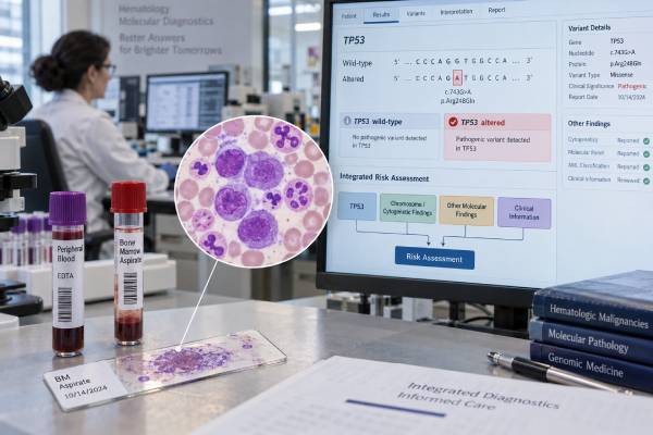
A TP53 mutation test looks for changes in TP53, a tumor suppressor gene that helps damaged cells stop dividing or undergo cell death. In acute myeloid leukemia (AML), myelodysplastic neoplasms (MDS), and related myeloid cancers, a pathogenic TP53 mutation often identifies biologically aggressive disease. The result can influence modern disease classification, prognostic risk, treatment planning, transplant discussions, and clinical-trial consideration. Interpretation is more complex than simply calling TP53 “positive” or “negative.” Clinicians also consider the variant allele frequency (VAF), whether one or both TP53 copies appear inactivated, chromosome findings such as a complex karyotype, blast percentage, other mutations, prior chemotherapy or radiation, and the patient’s overall clinical picture. A TP53 variant detected in blood is usually a somatic leukemia-related finding in this setting, but it does not by itself prove that the variant is inherited. Correct interpretation therefore depends on the specimen, assay, disease context, and the complete pathology report.
- A pathogenic TP53 mutation in AML or MDS is generally a high-risk finding associated with genomic instability, treatment resistance, and a greater chance of relapse.
- Allelic status matters: biallelic or “multi-hit” TP53 alteration usually carries more adverse biological significance than an isolated low-level finding.
- The result is not interpreted alone. Bone marrow morphology, cytogenetics, blast count, VAF, treatment history, and coexisting mutations all affect classification and prognosis.
- A blood or marrow TP53 mutation does not automatically mean Li-Fraumeni syndrome. Germline testing may require a carefully chosen non-hematopoietic specimen when hereditary disease is a concern.
- No single TP53 result determines treatment. The finding commonly prompts high-risk treatment planning, transplant assessment when appropriate, and consideration of clinical trials.
Table of Contents
- What a TP53 Mutation Test Measures
- Why TP53 Matters in Leukemia and MDS
- How to Read a Positive TP53 Result
- Classification and Prognostic Meaning
- Treatment and Transplant Implications
- Negative, VUS, and Low-Level Results
- Germline Versus Somatic TP53 Findings
What a TP53 Mutation Test Measures
TP53 encodes the p53 protein, an important safeguard against the survival and expansion of genetically damaged cells. When p53 function is lost, abnormal blood-forming cells can accumulate additional chromosome and DNA changes. This helps explain why TP53-altered myeloid cancers often have complex genomes and can be difficult to control with standard therapy.
In leukemia and MDS, TP53 is commonly tested as part of a next-generation sequencing (NGS) panel performed on bone marrow aspirate or peripheral blood. The test may detect single-nucleotide variants and small insertions or deletions. Depending on the laboratory, additional assays may help determine whether the second TP53 copy is deleted or altered. Chromosome analysis, fluorescence in situ hybridization, and copy-number methods can therefore provide information that sequencing alone may not show.
A molecular report usually lists the exact TP53 variant, its classification, and a variant allele frequency, or VAF. VAF is the percentage of sequencing reads carrying the variant. It is not the same as the percentage of leukemia cells. Tumor purity, normal-cell contamination, copy-number changes, loss of heterozygosity, and technical factors can all change the observed VAF.
TP53 testing is often performed alongside other molecular studies because AML and MDS are defined and risk-stratified by combinations of findings rather than by one gene alone. A broad leukemia molecular and MRD strategy may use different assays at diagnosis, during treatment, and after remission. TP53 NGS at diagnosis is primarily a classification and risk tool; it is not automatically an ideal marker for measurable residual disease in every patient.
The testing method also sets the limit of detection. A standard diagnostic panel may reliably detect variants only above a certain VAF, while specialized assays can detect much smaller clones. This matters when comparing results over time. A later “negative” result can mean that the clone fell below that assay’s detection threshold rather than that every TP53-mutated cell disappeared.
Why TP53 Matters in Leukemia and MDS
TP53 mutations occur in a minority of de novo AML and MDS cases but are enriched in biologically difficult settings. They are seen more often in older adults, in disease with complex chromosome abnormalities, and in myeloid neoplasms that develop after previous chemotherapy or radiation. TP53 alterations are also strongly associated with chromosome 5, chromosome 7, and other structural abnormalities in many high-risk cases.
The clinical importance of TP53 comes from the biology behind the result. A leukemia clone that has lost effective p53 signaling can tolerate DNA damage that would normally stop cell division. The clone may therefore continue evolving, acquire multiple chromosome abnormalities, and survive treatment-related stress. This pattern contributes to lower remission durability and a high relapse risk.
The strongest adverse signal is often seen when TP53 is biallelically inactivated, sometimes described as multi-hit TP53. That can occur through two pathogenic TP53 mutations or through one mutation combined with deletion, loss of heterozygosity, or another event that disables the remaining normal allele. A complex karyotype can support the interpretation that TP53 biology is driving broad genomic instability, although exact classification rules differ between current systems.
Not every TP53 mutation has identical meaning. A small TP53 clone at a low VAF may represent an emerging subclone, residual clonal hematopoiesis, or a component of the malignant clone. Conversely, a high-VAF mutation with chromosome 17p loss may strongly support biallelic inactivation. This is why the pathology team evaluates sequence data together with cytogenetics and morphology.
TP53 also differs from mutations that can define more favorable AML subsets. For example, NPM1 mutation testing in AML can carry different classification and monitoring implications. When multiple abnormalities coexist, current leukemia classification and risk frameworks—not a simple gene-by-gene ranking—should be used.
How to Read a Positive TP53 Result
A positive TP53 result usually means the laboratory identified a variant considered pathogenic or likely pathogenic. The most useful interpretation answers several additional questions.
First, what kind of variant was found? Many clinically important TP53 variants are missense substitutions within the DNA-binding domain, but nonsense, frameshift, splice-site, and other loss-of-function changes also occur. The exact molecular consequence may help a laboratory classify the variant, but routine clinical decisions are usually based more on whether the alteration is pathogenic, its allelic context, and the disease as a whole than on one specific amino-acid change.
Second, what is the VAF? A higher VAF can suggest that a large fraction of sampled cells carries the mutation, but it cannot by itself prove biallelic disease. A VAF near 50% may occur in a large somatic clone or in a constitutional germline variant. Values can also exceed 50% when the normal TP53 allele is lost. Very low values need careful interpretation because they may represent a small malignant clone, treatment-related clonal hematopoiesis, or technical uncertainty near the assay limit.
Third, is there evidence of a second TP53 hit? Reports may mention two TP53 mutations, 17p deletion, copy-neutral loss of heterozygosity, or “multi-hit” status. Some laboratories can infer allelic state from integrated NGS and copy-number data; others require separate cytogenetic or genomic testing. If allelic status is clinically important but not reported, the treating team may ask the laboratory how it was assessed.
Fourth, what else is present? Blast percentage, complex karyotype, antecedent MDS, prior cytotoxic therapy, and other myeloid mutations all influence the diagnosis. A TP53 finding should therefore be read in the context of the integrated hematopathology report, not as a standalone consumer genetic test.
Finally, the words pathogenic, likely pathogenic, and variant of uncertain significance (VUS) are not interchangeable. A VUS means the evidence is insufficient to call the change disease-causing or benign. A VUS should not be treated as if the patient definitely has TP53-mutated high-risk leukemia solely because the gene name appears on the report.
Classification and Prognostic Meaning
Modern myeloid classification places far more weight on genetics than older systems did. Two major frameworks published in 2022—the fifth edition World Health Organization (WHO) classification and the International Consensus Classification (ICC)—agree that TP53 biology is important, but they do not organize every TP53-mutated case in exactly the same way.
The ICC includes a family of myeloid neoplasms with mutated TP53, with categories spanning MDS, MDS/AML, and AML according to blast percentage and TP53 criteria. WHO-HAEM5 recognizes MDS with biallelic TP53 inactivation as a distinct MDS entity. In AML, WHO and ICC terminology can therefore differ even when they describe the same patient. A report that uses one system is not necessarily contradicting a report that uses the other.
For treatment decisions, classification is paired with prognostic systems. In the 2022 European LeukemiaNet (ELN) genetic risk framework for AML, pathogenic TP53 mutation is an adverse-risk feature under the framework’s specified criteria. The practical message is that a confirmed clinically significant TP53 mutation in AML usually signals a much greater risk of treatment failure and relapse than is seen in many TP53-wild-type AML subsets.
However, prognosis is not a fixed survival number. Published outcomes vary with age, fitness, disease stage, allelic status, cytogenetics, prior therapy, treatment regimen, response depth, and whether transplantation is possible. Older cohorts of TP53-mutated MDS/AML have shown very short median survivals, but quoting one number to an individual patient can be misleading. Newer supportive care, molecular selection, trial enrollment, and transplant strategies also change the population being treated.
A particularly important distinction is between a single low-level TP53 event and a multi-hit/biallelic pattern. Studies consistently show that allelic state and genomic complexity add prognostic information. The treating hematologist may therefore ask for copy-number or cytogenetic clarification rather than assuming every “TP53 positive” result carries exactly the same risk.
Response to treatment also changes the outlook. Achieving a morphologic remission, lowering the TP53 clone, and reaching transplant with controlled disease can be favorable signs, even though relapse risk remains substantial. Prognosis should consequently be updated over time rather than frozen at the day of diagnosis.
Treatment and Transplant Implications
TP53 testing does not identify a universally effective TP53-targeted drug for AML or MDS. Instead, the result mainly helps clinicians recognize a high-risk disease biology and choose an appropriate overall strategy.
For medically fit patients, intensive chemotherapy may be considered in selected circumstances, but TP53-mutated AML has historically shown lower durability of response than many other AML groups. For older or less-fit adults, hypomethylating agents such as azacitidine or decitabine, often combined with venetoclax in AML, are common treatment backbones. These regimens can produce remissions, but TP53-mutated disease frequently relapses and long-term disease control remains difficult.
This is an area where clinical-trial participation can be especially important. Several agents designed to exploit TP53-related vulnerabilities or immune pathways have been studied, yet promising early response rates have not always translated into successful randomized trials. A patient should therefore not assume that an experimental drug mentioned online is available, approved, or proven to improve survival. Trial eligibility and current evidence need to be checked at the time treatment is chosen.
Allogeneic hematopoietic cell transplantation is the main potentially curative approach for appropriate patients with TP53-mutated AML or high-risk MDS. Even after transplant, relapse rates are high. Outcomes tend to be better when disease is controlled before transplantation, so the decision weighs response, age, comorbidities, donor options, transplant risk, and patient goals. TP53 status can justify an early transplant consultation rather than waiting until multiple treatment failures.
The result may also affect the intensity of follow-up. Bone marrow evaluation, blood counts, cytogenetics, and molecular testing can be used to assess response. When a molecular marker is being followed, clinicians must understand whether persistence represents active leukemia, residual clonal hematopoiesis, or a clone whose significance is uncertain. A technically sensitive test is useful only when its result is clinically interpretable.
Treatment choices should also account for disease-defining abnormalities that may create separate therapeutic opportunities. A comprehensive leukemia workup can identify targetable mutations or fusions alongside TP53. The presence of TP53 does not erase the need to evaluate those findings, although it may still influence expected durability and overall risk.
Negative, VUS, and Low-Level Results
A negative TP53 test means no reportable TP53 alteration was detected by that assay in that specimen. It does not prove that p53 function is normal in every leukemia cell. The panel may not detect certain large deletions, structural changes, noncoding alterations, or very small clones. Poor specimen quality, low tumor content, or treatment before sampling can also reduce sensitivity.
A negative result also does not make AML or MDS low risk. Prognosis can be driven by other mutations, chromosome abnormalities, disease history, blast count, measurable residual disease, age, and treatment response. TP53 is one part of a larger molecular profile.
A VUS is different from a negative result, but it is also different from a pathogenic result. Laboratories classify variants using population frequency, functional evidence, known disease associations, computational data, and curated databases. As evidence grows, a VUS may later be reclassified. Until then, major leukemia decisions should not usually be based on the assumption that the VUS is functionally equivalent to a known pathogenic TP53 mutation.
Low-level pathogenic TP53 variants are another challenge. A small clone can be biologically meaningful, particularly if it fits the marrow and cytogenetic findings. But TP53 mutations also occur in age-related or therapy-related clonal hematopoiesis without meeting criteria for leukemia. After treatment, a persistent low-VAF TP53 clone may therefore require comparison with morphology, blood counts, cytogenetics, and the pretreatment molecular profile.
The most reliable question is not “Is TP53 present?” but “What does this specific TP53 finding mean in this specimen, at this time, in this disease?” That integrated approach prevents both overcalling a small incidental clone and underestimating a clearly high-risk multi-hit pattern.
Germline Versus Somatic TP53 Findings
Most TP53 mutations found during an AML or MDS workup are somatic, meaning they arose in the abnormal blood-cell clone. TP53 is also the major gene associated with Li-Fraumeni syndrome, an inherited cancer-predisposition condition. The overlap creates a practical testing problem: blood is an excellent specimen for leukemia sequencing but can be a poor specimen for deciding whether a TP53 variant is constitutional.
A VAF around 50% does not prove that a TP53 variant is germline. A large leukemia clone can produce a similar value. Conversely, a true constitutional variant can appear at a different VAF because the specimen contains abnormal clones or copy-number changes. TP53-mutated clonal hematopoiesis can also mimic a germline result, especially in older adults and people previously exposed to chemotherapy or radiation.
Germline evaluation may be considered when the personal or family history suggests a hereditary cancer syndrome, when cancer occurs unusually early, when multiple primary tumors are present, or when the molecular pattern raises concern. If testing is pursued, genetics professionals may recommend cultured skin fibroblasts or another validated non-hematopoietic source rather than relying only on peripheral blood or saliva, because saliva can contain blood-derived cells.
This distinction matters for relatives. A somatic leukemia mutation is not automatically inherited and generally does not imply a 50% risk to children. A confirmed germline pathogenic TP53 variant has completely different surveillance and family-testing implications. The separate TP53 testing context in other blood cancers also illustrates why the same gene can carry different disease-specific meanings.
The final interpretation should therefore state whether the laboratory tested tumor/blood cells only, whether germline origin was assessed, and whether genetic counseling is recommended. Patients should not infer Li-Fraumeni syndrome from a leukemia NGS report without appropriate confirmation.
References
- TP53-Mutated Myeloid Neoplasms: 2024 Update on Diagnosis, Risk-Stratification, and Management — 2025, review.
- TP53 mutations in myeloid neoplasms: implications for accurate laboratory detection, diagnosis, and treatment — 2024, review.
- TP53 in MDS and AML: Biological and clinical advances — 2024, review.
- A practical approach on the classifications of myeloid neoplasms and acute leukemia: WHO and ICC — 2024, review.
- TP53-Mutated Myelodysplastic Syndrome and Acute Myeloid Leukemia: Biology, Current Therapy, and Future Directions — 2022, review.
Disclaimer
This article is for general educational purposes and does not replace individualized diagnosis or treatment from a hematologist, hematopathologist, genetics professional, or transplant team. TP53 results must be interpreted with marrow findings, cytogenetics, treatment history, assay limitations, and the patient’s overall health. Treatment options and clinical-trial availability can change over time.





