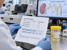
The transtubular potassium gradient (TTKG) is a calculated urine index historically used to estimate potassium secretion in the cortical collecting duct and to judge whether aldosterone is having an appropriate effect. It requires serum potassium, urine potassium, plasma osmolality, and urine osmolality. In hyperkalemia, a low TTKG has traditionally suggested inadequate distal potassium secretion from low aldosterone activity, aldosterone resistance, low distal sodium delivery, or impaired kidney function. In hypokalemia, a low TTKG can reflect appropriate potassium conservation. The calculation is physiologically appealing, but its core assumptions are imperfect, especially because solutes such as urea can be reabsorbed downstream in the medullary collecting duct. For that reason, TTKG has fallen out of favor in some modern nephrology practice. It can still provide context in selected patients, but it should not be used alone to diagnose hypoaldosteronism, adrenal disease, or the cause of a potassium disorder.
- TTKG estimates distal potassium secretion using urine potassium corrected for urine concentration relative to plasma.
- Traditional interpretation expects a higher TTKG during hyperkalemia and a lower TTKG during hypokalemia.
- A low TTKG in hyperkalemia can suggest impaired aldosterone effect, but kidney function and distal sodium delivery must be considered.
- The calculation is unreliable when urine is very dilute or when distal sodium delivery is inadequate, and its physiologic assumptions are debated.
- Severe hyperkalemia or hypokalemia is managed according to serum potassium, symptoms, ECG findings, and cause—not the TTKG.
Table of Contents
- What TTKG Measures and How It Is Calculated
- Traditional TTKG Interpretation
- TTKG, Aldosterone, and Hyperkalemia
- TTKG in Hypokalemia
- Why TTKG Has Important Limitations
- Alternatives to TTKG
- Collection, Follow-Up, and Safety
What TTKG Measures and How It Is Calculated
Potassium excretion is largely determined in the distal nephron. Principal cells in the connecting tubule and collecting duct reabsorb sodium and secrete potassium, a process strongly influenced by aldosterone, serum potassium, tubular flow, and distal sodium delivery.
The TTKG was designed to estimate the potassium concentration in fluid leaving the cortical collecting duct after correcting for water reabsorption. The common formula is:
TTKG = (urine potassium ÷ serum potassium) ÷ (urine osmolality ÷ plasma osmolality)
The result has no unit.
For example, if urine potassium is 40 mmol/L, serum potassium is 5 mmol/L, urine osmolality is 600 mOsm/kg, and plasma osmolality is 300 mOsm/kg:
TTKG = (40 ÷ 5) ÷ (600 ÷ 300) = 8 ÷ 2 = 4
The calculation assumes that the urine potassium concentration can be “back-corrected” for water removed after the cortical collecting duct. That allows the result to function as a rough index of the driving force for potassium secretion.
The related urine potassium test gives a simpler measurement of urinary potassium concentration. TTKG adds serum potassium and osmolality to make the number more physiologic, but that added complexity also creates more assumptions.
Traditional TTKG Interpretation
Classic teaching uses different expectations depending on the serum potassium.
| Clinical setting | Expected pattern | Traditional implication |
|---|---|---|
| Hypokalemia | Low TTKG, often <2–3 | Appropriate renal potassium conservation |
| Hyperkalemia | Higher TTKG, historically >7–10 | Appropriate distal potassium secretion |
| Hyperkalemia | Low TTKG, often <5–6 | Inadequate distal secretion; consider low aldosterone effect or other limiting factors |
These cutoffs are not universal modern reference ranges. They come from physiologic and older clinical literature and should be treated as rough guideposts.
A TTKG of 4 has a very different meaning in a patient with serum potassium of 2.8 mmol/L than in a patient with serum potassium of 6.2 mmol/L. In the first patient, reduced potassium secretion may be exactly what the kidney should do. In the second, it may be an insufficient response.
Before interpreting TTKG, clinicians should confirm that the potassium abnormality itself is real. Hemolyzed blood samples can cause pseudohyperkalemia. Severe hypokalemia may result from gastrointestinal loss, renal wasting, or a shift into cells. The serum potassium test and ECG determine urgency.
TTKG, Aldosterone, and Hyperkalemia
The most common traditional use of TTKG is evaluation of hyperkalemia.
Aldosterone promotes sodium reabsorption through epithelial sodium channels in the distal nephron and increases the electrochemical conditions that favor potassium secretion. If serum potassium is high, aldosterone secretion and renal potassium excretion should normally increase.
A low TTKG in hyperkalemia can therefore raise several possibilities:
- inadequate aldosterone production;
- reduced renin leading to low aldosterone;
- resistance to aldosterone action;
- medications that interfere with aldosterone signaling or epithelial sodium channels;
- insufficient distal sodium delivery;
- low tubular flow;
- advanced kidney dysfunction.
Diabetes with chronic kidney disease can produce hyporeninemic hypoaldosteronism, often associated with hyperkalemia and a mild normal-anion-gap metabolic acidosis. ACE inhibitors and angiotensin receptor blockers reduce aldosterone signaling. Spironolactone and eplerenone block the mineralocorticoid receptor. Amiloride and trimethoprim can reduce epithelial sodium channel activity. NSAIDs can reduce renin release in susceptible patients.
In these settings, TTKG cannot tell which mechanism is present. Direct renin and aldosterone measurements, kidney function, bicarbonate, medication review, and clinical volume status are more specific.
A urine anion gap or direct urine ammonium measurement may be relevant when hyperkalemia occurs with normal-anion-gap metabolic acidosis and type 4 renal tubular acidosis is being considered.
The fludrocortisone concept
Older teaching suggested repeating TTKG after fludrocortisone to distinguish aldosterone deficiency from resistance: a rise would support a hormone-deficiency state with preserved renal response. In modern practice, this approach is not routinely required and must be used cautiously because fludrocortisone can affect blood pressure, sodium balance, and fluid retention. Endocrine testing usually provides more direct information.
TTKG in Hypokalemia
When serum potassium is low, the kidney should reduce potassium secretion. A low TTKG can therefore support appropriate conservation, while an unexpectedly high value may suggest ongoing renal potassium wasting.
Renal potassium loss can result from:
- loop or thiazide diuretics;
- primary aldosteronism or other mineralocorticoid excess;
- Bartter or Gitelman syndromes;
- magnesium deficiency;
- some renal tubular acidoses;
- high distal sodium delivery and tubular flow.
However, TTKG is not the preferred first test in every hypokalemia workup. A spot urine potassium-to-creatinine ratio or 24-hour potassium excretion often provides a simpler estimate of whether the kidneys are losing potassium.
The fractional excretion of potassium (FEK) is another creatinine-corrected index. It does not rely on urine osmolality, although it has its own limitations and lacks one universal threshold for all clinical settings.
Acid-base status helps narrow the differential. Hypokalemia with metabolic alkalosis and high urine chloride suggests a different process than hypokalemia with metabolic acidosis. Blood pressure provides another key split: hypertension makes mineralocorticoid excess more likely, while normal or low blood pressure is more compatible with diuretic effects or salt-wasting tubulopathies.
Magnesium should be measured because hypomagnesemia can cause persistent renal potassium wasting. Potassium replacement may be ineffective until magnesium deficiency is corrected.
Why TTKG Has Important Limitations
TTKG depends on assumptions that are not always valid.
Urine should not be dilute
The formula assumes water is being reabsorbed downstream so that urine osmolality can correct the potassium concentration back toward the cortical collecting duct value. If urine osmolality is lower than plasma osmolality, that assumption breaks down. TTKG is generally considered uninterpretable in a water diuresis.
Distal sodium delivery must be adequate
Potassium secretion requires sodium delivery to the distal nephron. Historically, a urine sodium above roughly 25 mmol/L was suggested as a condition for interpreting TTKG. If urine sodium is very low, a low TTKG may reflect lack of sodium delivery rather than low aldosterone activity.
Urea is not inert downstream
One of the most important criticisms is that the medullary collecting duct does not simply remove water while leaving solute unchanged. Urea is actively involved in medullary recycling and can be reabsorbed. This violates a core simplifying assumption used in the TTKG correction.
Potassium can also be handled downstream
The original model assumes little potassium transport after the cortical collecting duct. Under severe potassium depletion or other physiologic conditions, downstream potassium handling can change.
CKD and low urine flow complicate interpretation
Advanced kidney disease, oliguria, and altered tubular architecture change potassium handling. The kidney’s ability to excrete potassium depends on GFR, remaining nephron adaptation, distal sodium delivery, and flow. A single TTKG cannot separate these effects.
For these reasons, contemporary reviews often describe TTKG as an adjunct that has fallen out of favor rather than a definitive aldosterone test.
Alternatives to TTKG
Several simpler measurements can answer related questions.
Spot urine potassium-to-creatinine ratio adjusts urine potassium for sample concentration without requiring urine osmolality. In hypokalemia, a low ratio supports appropriate renal conservation; a high ratio suggests renal wasting. Exact thresholds depend on units and context.
24-hour urine potassium directly estimates daily excretion. It is useful when intake and losses are stable, but acute potassium disorders can change faster than a 24-hour collection captures.
Fractional excretion of potassium uses urine and serum potassium plus urine and serum creatinine. A recent 2026 study in patients with hypoaldosteronism found that FEK and other urinary ratios correlated with TTKG and may serve as practical alternatives.
Renin and aldosterone are more directly related to the hormonal question. Their interpretation requires attention to posture, sodium intake, medications, potassium concentration, and assay conditions, but they can distinguish low hormone production from some other mechanisms more directly than TTKG.
Serum bicarbonate and acid-base testing help identify type 4 renal tubular acidosis, where hypoaldosteronism or aldosterone resistance contributes to hyperkalemia and impaired ammonium excretion. The bicarbonate blood test is a simple starting point.
No single urine index is sufficient. Modern evaluation usually favors a pattern-based approach using serum potassium, kidney function, acid-base status, urine potassium excretion, urine sodium, blood pressure, and medications.
Collection, Follow-Up, and Safety
TTKG requires a spot urine potassium and osmolality together with serum potassium and plasma or serum osmolality collected at roughly the same time. A serum sodium and kidney function panel are usually available as part of the same evaluation.
Do not intentionally alter fluid intake or stop medications to manipulate the result unless a clinician gives specific instructions. Diuretics, renin-angiotensin system blockers, mineralocorticoid receptor antagonists, potassium supplements, potassium binders, trimethoprim, NSAIDs, and IV fluids can all affect interpretation.
If TTKG is unexpectedly low in hyperkalemia, the next steps may include confirming serum potassium, reviewing medications, measuring creatinine and bicarbonate, assessing urine output, and checking renin, aldosterone, and cortisol-related testing when indicated. If kidney function is substantially reduced, diminished excretory capacity may be a major part of the explanation.
If TTKG is unexpectedly high in hypokalemia, clinicians often look for diuretic exposure, magnesium deficiency, mineralocorticoid excess, renal tubular disorders, and acid-base abnormalities.
Urgency depends on the serum potassium, not the TTKG. Palpitations, syncope, severe weakness, paralysis, chest symptoms, or a markedly high or low potassium level warrant urgent assessment. Hyperkalemia can cause life-threatening arrhythmias even before obvious symptoms appear.
TTKG can still be educational because it forces attention to distal potassium secretion and aldosterone physiology. Clinically, its best role is narrow: an optional supporting calculation when its prerequisites are met and when it agrees with more direct evidence.
Practical examples: when TTKG agrees or conflicts with the clinical picture
Hyperkalemia with low TTKG and adequate urine sodium: Suppose serum potassium is 6.0 mmol/L, urine is more concentrated than plasma, urine sodium is adequate, and TTKG is 3. Under the traditional model, the distal nephron is not increasing potassium secretion as much as expected. Hypoaldosteronism, aldosterone resistance, or a drug that blocks distal sodium uptake becomes plausible. The next step is not to diagnose the cause from TTKG, but to review medications, kidney function, acid-base status, and—when appropriate—renin and aldosterone.
Hyperkalemia with low TTKG and very low urine sodium: Here the traditional calculation is much less informative. The distal nephron may simply lack sodium delivery, so potassium secretion cannot proceed normally even if aldosterone is present. Severe volume depletion, low effective circulating volume, or advanced kidney dysfunction can produce this situation. Treating the TTKG as proof of aldosterone deficiency would be a mistake.
Hypokalemia with a high TTKG: If serum potassium is 2.8 mmol/L and the TTKG remains high while urine is appropriately concentrated, renal potassium wasting may be present. Diuretics, mineralocorticoid excess, Bartter or Gitelman physiology, or magnesium deficiency are possibilities. A spot urine potassium-to-creatinine ratio, blood pressure, chloride, bicarbonate, and magnesium often provide more directly useful information.
Dilute urine: If urine osmolality is below plasma osmolality, TTKG should generally not be interpreted. The correction for water reabsorption no longer represents the process the formula assumes. A numerical result can still be calculated, but that does not make it physiologically valid.
Why many clinicians now favor simpler urinary potassium indices
The TTKG model assumed that little potassium was added to or removed from the terminal collecting duct after the cortical collecting duct and that urine osmolality could reliably correct for downstream water reabsorption. Later physiologic work showed that urea recycling and medullary handling make those assumptions imperfect. As a result, many nephrologists no longer use TTKG as a routine stand-alone test.
Recent research has evaluated alternatives such as FEK and spot urine potassium-to-creatinine ratios in suspected hypoaldosteronism. These indices also have limitations, but they do not depend on the same osmolality correction. No urinary index can fully replace the physiologic context.
Aldosterone testing still requires context
Renin and aldosterone concentrations are influenced by posture, sodium intake, potassium concentration, blood pressure medications, diuretics, kidney function, and time of day. In hyperkalemia, aldosterone should be interpreted relative to the stimulus: a value that falls within a laboratory’s population reference range may still be inappropriately low for a markedly elevated potassium.
This same principle explains the best use of TTKG. The question is not whether the number is inside a generic “normal range.” The question is whether renal potassium secretion is appropriate for the serum potassium, distal sodium delivery, urine flow, and hormonal setting. When those prerequisites are not met, a different urine potassium measure and direct evaluation of the suspected cause are usually more informative.
Interpreting a TTKG report from the laboratory
Some laboratories calculate TTKG automatically, while others provide only the four values needed for the formula. Before accepting the number, confirm that serum and urine samples were obtained close together and that urine osmolality is at least as high as plasma osmolality. Also check urine sodium and urine flow if available.
A mathematically correct TTKG can still be physiologically uninterpretable when these conditions are not met. This is an important distinction: the formula will always produce a number, but the body may not be in the state assumed by the model.
If an older chart or consultation note cites a TTKG threshold, it is reasonable to reinterpret that result with modern caution. Current practice increasingly favors direct assessment of serum potassium, kidney function, urine potassium indices, medications, acid-base status, and renin-aldosterone physiology rather than using one TTKG cutoff as decisive evidence.
A normal-looking TTKG also should not delay treatment of a dangerous potassium abnormality. Hyperkalemia management is based on the serum potassium level, ECG findings, symptoms, rate of change, and the underlying cause. The urine calculation can be revisited after immediate safety issues are addressed. In hypokalemia, severe weakness, arrhythmia risk, or marked potassium depletion likewise takes priority over refining a urinary index.
References
- Beyond the transtubular potassium gradient: Alternative urinary markers for diagnosing hypoaldosteronism 2026
- Clinical manifestations and associated factors in acquired hypoaldosteronism in endocrinological practice 2022
- Hyperkalemia in Chronic Kidney Disease: Links, Risks and Management 2022 (Review)
- Pathophysiology and clinical management of hyperkalemia in chronic kidney disease 2023 (Review)
- Extrarenal Effects of Aldosterone on Potassium Homeostasis 2022 (Review)
Disclaimer
TTKG is an older adjunctive index with important physiologic limitations and should not be used alone to diagnose aldosterone deficiency, aldosterone resistance, or the cause of a potassium disorder. Severe potassium abnormalities can be life-threatening and require assessment based on serum potassium, ECG findings, symptoms, kidney function, and the underlying cause.