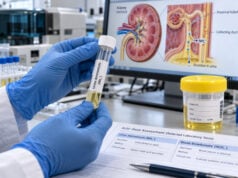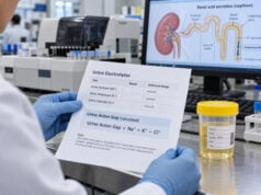
Fractional excretion of uric acid (FEUA) estimates what percentage of urate filtered by the kidneys is excreted in urine. It combines paired blood and urine uric acid and creatinine measurements and is mainly used to understand renal urate handling. A low FEUA suggests that the kidneys are retaining a larger share of filtered urate, a common mechanism in hyperuricemia and gout. A high FEUA suggests increased renal urate loss and can occur with renal hypouricemia, proximal tubular disorders, some medications, and several causes of hyponatremia. FEUA is also used in selected patients to help distinguish syndrome of inappropriate antidiuresis (SIAD) from other fluid-balance disorders. There is no single FEUA cutoff that diagnoses gout or any one kidney disorder. Interpretation depends on serum uric acid, kidney function, volume status, sodium, medications, diet, and whether the patient is taking urate-lowering or uricosuric drugs.
- FEUA shows the percentage of filtered urate excreted by the kidneys and requires paired serum and urine uric acid and creatinine.
- A low FEUA supports renal urate underexcretion, a common contributor to hyperuricemia and gout.
- A high FEUA supports renal urate wasting and may occur in renal hypouricemia, proximal tubule injury, SIAD, or with uricosuric drugs.
- Many healthy adults have FEUA values in roughly the mid-single digits to low teens, but laboratory and clinical context matter more than one universal range.
- FEUA does not replace serum uric acid, kidney function tests, stone evaluation, or crystal confirmation when gout diagnosis is uncertain.
Table of Contents
- What FEUA Measures and How It Is Calculated
- Normal, High, and Low FEUA
- FEUA in Gout and Hyperuricemia
- High FEUA, Hypouricemia, and Tubular Disorders
- FEUA in Hyponatremia and SIAD
- Collection, Medications, and Confounders
- Follow-Up and Related Tests
What FEUA Measures and How It Is Calculated
Uric acid is the end product of purine metabolism in humans. In blood, most exists as urate. The kidneys filter urate freely at the glomerulus, then the proximal tubule reabsorbs and secretes it through several transporters. The net result is that most filtered urate is reabsorbed and only a minority is excreted.
The standard calculation is:
FEUA (%) = (urine uric acid × serum creatinine) ÷ (serum uric acid × urine creatinine) × 100
Urine and blood samples should be collected close together. Serum and urine uric acid must use corresponding units, and serum and urine creatinine must use corresponding units.
FEUA differs from a 24-hour urine uric acid measurement. The 24-hour test asks how much uric acid leaves the body in a day. FEUA asks what fraction of the urate presented to the kidney through filtration reaches the urine. The two can point in the same direction but answer different questions.
For example, a person with high serum uric acid and low FEUA may have renal urate underexcretion. Another person may have a high total 24-hour uric acid excretion because production and dietary purine load are high even if the fractional excretion is not unusually high.
The uric acid blood test is therefore the starting point. FEUA becomes useful when the clinician wants to know why serum urate is high or low, or whether the kidney is handling urate appropriately.
Normal, High, and Low FEUA
There is no single universally accepted reference interval across all laboratories and clinical settings. In adults with normal kidney function, FEUA is often roughly in the 5%–12% range. Sex, serum urate, kidney function, sodium balance, medications, and genetics can shift the value.
| Pattern | What it suggests | Examples |
|---|---|---|
| Low FEUA | Increased net renal urate reabsorption or reduced urate secretion | Volume depletion, many cases of gout/hyperuricemia, some drug effects |
| Mid-range FEUA | Typical net urate handling | Normal physiology; interpretation still depends on serum urate |
| High FEUA | Increased renal urate loss | Renal hypouricemia, Fanconi syndrome, SIAD, uricosuric drugs, some tubular injuries |
A “normal” FEUA does not guarantee normal uric acid balance. Uric acid levels reflect production, intestinal excretion, filtration, and multiple tubular transport processes. Kidney function also changes the filtered load.
Several urate transporters help explain the patterns. URAT1 and GLUT9 are major reabsorptive pathways, while other transporters contribute to secretion. Genetic changes affecting these proteins can cause unusually low serum urate with excessive urinary urate loss or contribute to hyperuricemia.
The kidney normally handles urate dynamically. Sodium and urate reabsorption are linked in the proximal tubule, so volume contraction tends to lower urate excretion while volume expansion can increase it.
FEUA in Gout and Hyperuricemia
Most hyperuricemia is related more to impaired excretion than to massive uric acid overproduction. That does not mean every person with gout needs FEUA testing.
Gout is caused by deposition of monosodium urate crystals in joints and tissues. Serum urate helps assess the biochemical environment, but a normal serum urate during an acute flare does not exclude gout. When the diagnosis is uncertain, crystal identification from joint fluid is more direct than FEUA.
FEUA may help characterize the mechanism of persistent hyperuricemia. A low FEUA supports renal underexcretion. This can be influenced by:
- chronic kidney disease;
- volume depletion;
- obesity and metabolic factors;
- genetic variation in urate transporters;
- thiazide and loop diuretics;
- low-dose aspirin;
- cyclosporine and some other drugs.
A urine uric acid test or 24-hour urine uric acid measurement may be more useful when evaluating stone risk or total daily uric acid excretion.
FEUA is not generally used to decide the target serum urate during gout treatment. Treatment-to-target strategies rely on serum uric acid measurements and the clinical course. FEUA can, however, help explain why the serum level behaves as it does and may be relevant when selecting or understanding uricosuric therapy.
Underexcretion does not mean the kidneys are “failing”
A low FEUA can occur in people with normal filtration. It reflects tubular handling, not just glomerular function. Conversely, chronic kidney disease can raise serum urate because total excretory capacity falls even if remaining nephrons alter fractional excretion adaptively.
The eGFR test and serum creatinine are required to assess overall filtration.
High FEUA, Hypouricemia, and Tubular Disorders
A high FEUA is especially useful when serum uric acid is unexpectedly low. The combination suggests that the kidneys are losing urate.
Renal hypouricemia is a rare inherited condition often caused by impaired urate reabsorption through URAT1 or GLUT9. Affected people can have very low serum uric acid and markedly increased fractional urate excretion. Many are asymptomatic, but exercise-induced acute kidney injury and uric acid stones can occur.
Fanconi syndrome causes generalized proximal tubular dysfunction. In addition to urate wasting, patients may lose glucose despite normal blood glucose, phosphate, bicarbonate, amino acids, and other filtered substances. A high FEUA in this setting is one clue among several.
Acquired proximal tubular injury can occur with medications, toxins, monoclonal protein disorders, or other diseases. The pattern is broader than isolated urate loss.
Uricosuric medications intentionally increase urinary uric acid excretion by reducing reabsorption. This can raise FEUA. Losartan and some SGLT2 inhibitors can also lower serum urate through renal mechanisms, although their effects differ from dedicated uricosuric drugs.
A high FEUA should therefore trigger a medication review before a rare inherited diagnosis is considered.
Kidney stone implications
High urinary uric acid concentration can contribute to stones, especially when urine is persistently acidic and volume is low. However, FEUA alone does not quantify stone risk. Urine pH, total 24-hour uric acid, urine volume, citrate, sodium, calcium, and other factors matter. A 24-hour urine kidney stone risk test is more appropriate for recurrent stone evaluation.
FEUA in Hyponatremia and SIAD
FEUA has a second important use outside gout: evaluating some patients with hypotonic hyponatremia.
SIAD causes water retention with inappropriately concentrated urine. Mild extracellular volume expansion alters proximal tubular handling and can increase urate excretion, producing low serum uric acid and elevated FEUA. Values above roughly 10%–12% have often been associated with SIAD in appropriate clinical settings.
This is not a stand-alone SIAD test. The diagnosis still requires confirmation of hypotonic hyponatremia, inappropriately concentrated urine, exclusion of major thyroid and adrenal causes, and assessment of kidney function and diuretic use.
FEUA may be particularly helpful when urine sodium is hard to interpret because a patient is taking a diuretic. It has also been studied in attempts to distinguish SIAD from renal or cerebral salt wasting. One proposed clue is what happens after serum sodium is corrected: FEUA often normalizes in SIAD, whereas persistent elevation may support ongoing renal salt wasting. This distinction remains clinically difficult and should not be made from FEUA alone.
For the basic fluid-balance evaluation, clinicians commonly begin with urine osmolality and urine sodium. FEUA is an additional tool when the initial pattern is ambiguous.
Volume depletion usually has the opposite effect. Proximal sodium and urate reabsorption increase, serum uric acid can rise, and FEUA may fall.
Collection, Medications, and Confounders
FEUA is typically calculated from a spot urine sample collected near a blood sample. No special fasting is always required, but the purpose of the test matters.
Before interpretation, review:
- allopurinol or febuxostat, which reduce uric acid production;
- probenecid or other uricosuric drugs;
- diuretics;
- losartan;
- SGLT2 inhibitors;
- low-dose aspirin;
- cyclosporine or tacrolimus;
- recent IV fluids or major changes in sodium intake;
- kidney function and urine output.
Diet can affect urate production. High purine intake, alcohol, and fructose exposure can raise uric acid, while fasting and ketosis can alter urate handling. A one-time FEUA result during an acute illness may not represent a person’s usual state.
Creatinine-based calculations are also affected by rapidly changing kidney function. During AKI, serum creatinine is not at steady state, so fractional excretion values require caution.
A separate FENa test may be useful if volume status or AKI is the main question, while FEUA is more specific to urate handling.
Follow-Up and Related Tests
Follow-up depends on whether FEUA is being used for high uric acid, low uric acid, hyponatremia, or suspected tubular disease.
For persistent hyperuricemia or gout, useful next steps may include repeated serum urate, kidney function testing, medication review, and assessment for stones. FEUA can explain underexcretion, but it does not replace gout treatment decisions based on symptoms, flare history, tophi, serum urate, comorbidity, and kidney function.
For low serum uric acid with high FEUA, clinicians may check urine glucose, phosphate, bicarbonate, amino acids, and protein patterns for proximal tubular dysfunction. Very low uric acid with a compatible history can lead to testing for inherited renal hypouricemia.
For hyponatremia, serum osmolality, urine osmolality, urine sodium, glucose, cortisol-related evaluation, and thyroid testing are generally more fundamental. Severe symptomatic hyponatremia—with confusion, seizures, profound drowsiness, or other neurologic symptoms—is a medical emergency.
For kidney stones, a uric acid stone risk test focuses on urine pH, volume, and uric acid burden rather than FEUA alone.
FEUA is most useful as a mechanistic test. It tells whether the kidney is retaining or losing an unusually large fraction of filtered urate. The diagnosis comes from combining that information with the serum uric acid level and the clinical problem.
Practical FEUA interpretation patterns
FEUA becomes much easier to understand when it is paired with the serum uric acid concentration.
High serum uric acid with low FEUA: This is the classic underexcretion pattern. The kidneys are clearing a relatively small fraction of filtered urate, which can contribute to hyperuricemia and gout. Common influences include kidney dysfunction, volume depletion, insulin resistance, and medications that favor urate retention. This pattern describes a mechanism; it does not by itself establish gout, which is a crystal disease diagnosed from the clinical presentation and, when needed, joint-fluid analysis.
High serum uric acid with higher FEUA: The kidneys may be excreting a substantial fraction of urate, suggesting that increased urate production or a large filtered load is contributing. High cell turnover, tumor lysis, some hematologic disorders, and major purine loads can produce this pattern. The absolute urine uric acid excretion and clinical context are useful because a fractional percentage alone does not measure total uric acid production.
Low serum uric acid with high FEUA: This pattern points toward inappropriate renal urate wasting. In marked hypouricemia, inherited renal hypouricemia due to urate-transporter defects is one possibility. Generalized proximal tubular dysfunction can also cause high FEUA together with glucosuria despite normal blood glucose, phosphate wasting, bicarbonate loss, aminoaciduria, or low-molecular-weight proteinuria.
Hyponatremia with elevated FEUA: In hypotonic hyponatremia, an FEUA above roughly 10%–12% can support SIAD in the right setting, particularly when serum uric acid is low. However, cerebral or renal salt-wasting states may show a similar initial pattern. Some diagnostic approaches look at whether FEUA normalizes after the sodium disorder is corrected: normalization can fit SIAD, while persistent elevation may favor ongoing renal salt wasting. This approach requires careful specialist interpretation.
Treatment can change FEUA substantially
Uricosuric medicines intentionally increase urate excretion and FEUA. Losartan and SGLT2 inhibitors can also increase urinary urate to varying degrees. Diuretics often increase serum uric acid through volume and tubular effects, but the measured FEUA depends on timing and the individual’s kidney function. Xanthine oxidase inhibitors such as allopurinol reduce uric acid production, changing the filtered load and sometimes making comparisons with pretreatment values difficult.
For this reason, an isolated FEUA should be interpreted alongside a complete medication list and a serum uric acid obtained at about the same time.
FEUA does not equal daily uric acid excretion
A fractional excretion calculation answers what percentage of filtered urate appears in urine. A 24-hour urine uric acid measurement answers how much uric acid is excreted over a day. Those are related but not interchangeable questions.
Someone with a reduced GFR can have a particular FEUA percentage but a lower total filtered urate load. Conversely, a person with high urate production may excrete a large absolute amount even if the fractional percentage is not strikingly high. For gout and kidney stone evaluation, clinicians may therefore use both serum urate and timed urine uric acid when the distinction between overproduction, underexcretion, and stone risk would change management.
The most reliable interpretation of FEUA combines serum uric acid, creatinine, kidney function, sodium and volume status, medications, and the reason the test was ordered. A percentage outside a customary range is a clue to renal handling, not a stand-alone diagnosis.
When repeating FEUA is useful
A repeat FEUA can be valuable when the first sample was collected during dehydration, acute illness, changing kidney function, or after a medication was recently started. The goal is not to chase a specific percentage but to see whether the renal-handling pattern persists once the patient is clinically stable.
In hyponatremia, repeat testing after correction may sometimes help distinguish SIAD from persistent renal salt-wasting physiology, but this is a specialist use and must be interpreted with the sodium trajectory and treatment. In suspected inherited renal hypouricemia, a consistently very low serum uric acid level with persistently high FEUA is more convincing than one isolated sample.
For gout, repeating FEUA is usually less important than following serum urate to the treatment target and monitoring symptoms. FEUA can explain why urate is high, but it generally does not replace outcome-focused monitoring once treatment decisions have been made.
References
- Role of urate transporters in the kidneys and intestine in uric acid homeostasis 2026 (Review)
- Uric acid transport, transporters, and their pharmacological targeting 2023 (Review)
- Altered Serum Uric Acid Levels in Kidney Disorders 2022 (Review)
- Syndrome of Inappropriate Antidiuresis: From Pathophysiology to Management 2023 (Review)
- Gout associated with reduced renal excretion of uric acid. Renal tubular disorder that nephrologists do not treat 2022 (Review)
- Clinical practice guideline for renal hypouricemia (1st edition) 2019 (Guideline)
Disclaimer
FEUA is an interpretive calculation, not a stand-alone diagnosis of gout, SIAD, renal hypouricemia, or kidney disease. Results can be changed by kidney function, volume status, diet, and medications. Do not alter urate-lowering, diuretic, or fluid therapy based only on FEUA without guidance from a qualified clinician.