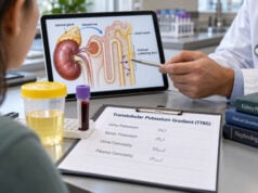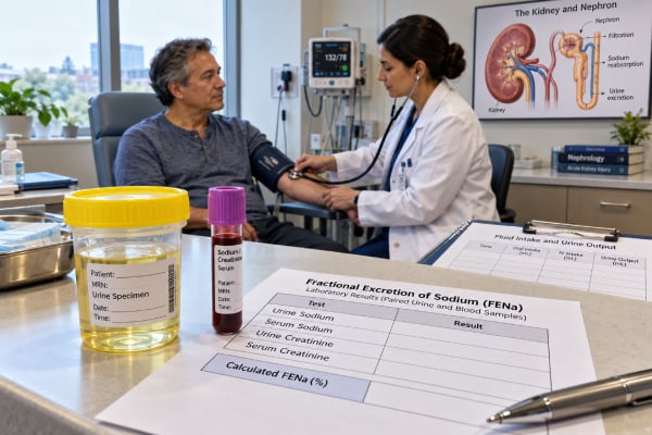
Fractional excretion of sodium (FENa) estimates the percentage of filtered sodium that the kidneys excrete in urine. It is calculated from paired urine and blood sodium and creatinine measurements and is used mainly during acute kidney injury (AKI). A low FENa can support a sodium-conserving, “prerenal” pattern, while a higher FENa can support tubular injury, but the calculation is not a stand-alone test for dehydration or for the cause of AKI. Diuretics, chronic kidney disease, sepsis, contrast exposure, glomerular disease, obstruction, and several other conditions can produce misleading results. FENa is most useful in an oliguric patient with newly increased creatinine who is not taking diuretics and does not have important pre-existing kidney disease. Even then, clinicians combine it with history, blood pressure, fluid status, urine output, urinalysis and sediment, medications, imaging when needed, and the trend in serum creatinine.
- FENa is calculated from simultaneous urine sodium, urine creatinine, serum sodium, and serum creatinine.
- A FENa below about 1% often supports sodium conservation, but it does not prove dehydration or reversible prerenal AKI.
- A FENa above about 2% may support tubular injury, but many intrinsic kidney diseases can still have a low FENa.
- Diuretics and chronic kidney disease reduce diagnostic accuracy, so FENa should be interpreted cautiously in those settings.
- AKI with very low urine output, breathing difficulty, severe swelling, confusion, or dangerous potassium changes needs prompt medical evaluation.
Table of Contents
- What FENa Is and How It Is Calculated
- FENa Cutoffs and What They Mean
- When FENa Is Most Useful
- When FENa Can Mislead
- FENa Versus FEUrea and Other AKI Tests
- Collection, Calculation, and Practical Examples
- Follow-Up and Urgent Findings
What FENa Is and How It Is Calculated
FENa asks how much of the sodium filtered through the glomeruli reaches the final urine. The usual formula is:
FENa (%) = (urine sodium × serum creatinine) ÷ (serum sodium × urine creatinine) × 100
The urine and serum samples should be collected at approximately the same time. Because serum creatinine can change quickly during AKI, using values from different time points can distort the calculation.
The physiologic idea is straightforward. When kidney perfusion or effective circulating volume falls but the tubules are still functioning, the kidneys activate sodium-retaining pathways. Urine sodium often falls and FENa may become very low. In classic acute tubular injury, damaged tubules may be less able to reabsorb filtered sodium, so urine sodium and FENa can rise.
That model is useful, but real AKI is more complicated. “Prerenal” and “intrinsic” are not always cleanly separated states. Sepsis, heart failure, cirrhosis, glomerular disease, obstruction, and drug effects can alter filtration, tubular handling, and neurohormonal signaling at the same time.
FENa also does not measure total body sodium or total body water. A person with heart failure may have excess body sodium and edema while the kidneys still avidly conserve sodium because effective arterial blood volume is low. A separate urine sodium test can provide related information, but it has the same need for clinical context.
FENa Cutoffs and What They Mean
Traditional teaching uses three broad zones:
| FENa | Typical interpretation | Important limitation |
|---|---|---|
| <1% | Sodium-conserving pattern; often seen in prerenal AKI | Can also occur in several intrinsic kidney diseases |
| 1%–2% | Intermediate or indeterminate | Overlap is substantial |
| >2% | Reduced tubular sodium reabsorption; can support acute tubular injury | Diuretics and CKD can raise FENa without acute tubular necrosis |
These are clinical heuristics, not diagnostic borders. A 2022 systematic review found that using a 1% threshold performed best in carefully selected oliguric patients without chronic kidney disease or diuretic exposure. Diagnostic performance was materially worse when those confounders were present.
A FENa below 1% therefore means “the kidney is excreting a small fraction of filtered sodium,” not “the patient is definitely dehydrated.” The cause may be true volume loss from vomiting, diarrhea, bleeding, or poor intake, but it can also be heart failure, cirrhosis, early obstruction, or some forms of intrinsic kidney disease.
Likewise, a FENa above 2% means sodium reabsorption is relatively reduced. This can be compatible with acute tubular injury, but a recent loop diuretic dose can produce the same direction of change by intentionally blocking sodium reabsorption.
The acute kidney injury blood test panel and urine output establish whether AKI is present and how severe it may be. FENa is a secondary etiologic clue.
When FENa Is Most Useful
FENa has its highest diagnostic value when the patient resembles the population in which the classic physiology applies.
The most favorable setting is:
- acute rise in serum creatinine;
- oliguria, meaning reduced urine output;
- no recent loop or thiazide diuretic;
- no substantial chronic kidney disease;
- no obvious glomerular, interstitial, vascular, or obstructive kidney process;
- paired urine and blood samples obtained before major treatment changes.
In that setting, a very low FENa can strengthen the impression that intact tubules are conserving sodium because kidney perfusion is reduced. A higher FENa can strengthen concern for tubular injury.
The calculation can be especially useful when the physical examination is unclear. Clinical estimates of volume status are imperfect. Dry mucous membranes, edema, blood pressure, heart rate, jugular venous pressure, weight change, and orthostatic symptoms each provide pieces of information but do not independently settle the diagnosis.
FENa may also be useful as part of a broader fluid and electrolyte evaluation. A fluid balance urine panel can combine urine sodium with osmolality and creatinine to show whether the kidneys are conserving water and salt appropriately.
FENa is a response marker, not a kidney-injury biomarker
FENa describes sodium handling. It does not directly measure tubular cell damage. A patient can have structural injury with a low FENa, especially early in the course or in diseases that do not primarily impair sodium reabsorption. Conversely, a patient can have an elevated FENa from medication without meaningful tubular necrosis.
That distinction matters because terms such as “prerenal” and “acute tubular necrosis” can imply certainty that the test does not provide.
When FENa Can Mislead
Several common situations break the simple low-versus-high interpretation.
Diuretics
Loop and thiazide diuretics increase sodium excretion. They can raise urine sodium and FENa even when the patient has low effective circulating volume. This is one reason clinicians sometimes calculate fractional excretion of urea instead.
Chronic kidney disease
CKD can reduce sodium reabsorptive reserve and alter baseline fractional excretion. A person with fewer functioning nephrons may need each remaining nephron to excrete a larger fraction of its filtered sodium to maintain balance. A FENa above 1% is therefore less specific for acute tubular injury in CKD.
Sepsis
Sepsis-associated AKI does not always follow the classic “low perfusion, then tubular necrosis” sequence. Hemodynamic changes, inflammation, microcirculatory dysfunction, and tubular responses can coexist. A low FENa is common in sepsis and does not exclude intrinsic injury.
Glomerular and vascular disease
Acute glomerulonephritis, vasculitis, and some vascular kidney disorders can present with low FENa because tubular sodium reabsorption remains active. Urinalysis and microscopy may provide much more important clues, such as blood, protein, dysmorphic red cells, or casts. A urine microscopy evaluation can therefore be decisive.
Obstruction
Early urinary obstruction can sometimes produce a low FENa. Later, impaired tubular function may increase it. Imaging is required when obstruction is plausible; FENa cannot rule it out.
Contrast, pigment injury, and other exceptions
Contrast-associated AKI, rhabdomyolysis, and some drug-related injuries may show low FENa despite intrinsic kidney injury. A urine myoglobin test or creatine kinase measurement may be relevant when muscle breakdown is suspected.
Because exceptions are common, FENa should modify a clinical probability rather than replace the differential diagnosis.
FENa Versus FEUrea and Other AKI Tests
Fractional excretion of urea, or FEUrea, was developed partly because proximal urea handling was thought to be less affected by diuretics than sodium handling. A value below about 35% has traditionally been used to support prerenal physiology.
However, modern evidence shows that FEUrea is not a perfect rescue test. A 2024 systematic review and meta-analysis found only modest diagnostic performance and did not establish FEUrea as clearly superior to FENa, even among patients receiving diuretics. The related FEUrea test is therefore best viewed as another piece of evidence.
Other AKI tools include:
- serum creatinine trend: confirms changing filtration but rises after injury has occurred;
- urine output: sensitive to changes in kidney function and part of AKI staging;
- urinalysis and sediment: can identify glomerular disease, interstitial inflammation, casts, or pigment;
- renal ultrasound: evaluates obstruction and kidney structure;
- medication and hemodynamic review: often reveals the cause more directly than a fractional excretion calculation;
- newer tubular injury biomarkers: useful in selected settings but not universally required.
FENa performs best when it answers a narrow question inside this broader evaluation: Is the kidney avidly conserving sodium right now?
Collection, Calculation, and Practical Examples
No special diet or fasting is usually required for an acute FENa. The important issue is timing. Ideally, collect blood and urine before IV saline, diuretics, or other interventions when doing so does not delay necessary care.
Suppose urine sodium is 15 mmol/L, serum sodium is 140 mmol/L, urine creatinine is 100 mg/dL, and serum creatinine is 2.0 mg/dL:
FENa = (15 × 2.0) ÷ (140 × 100) × 100 = 0.21%
That is a strongly sodium-conserving pattern. It supports prerenal physiology if the rest of the clinical picture fits, but it does not prove simple dehydration.
Now suppose urine sodium is 70 mmol/L, serum sodium 138 mmol/L, urine creatinine 50 mg/dL, and serum creatinine 2.0 mg/dL:
FENa = (70 × 2.0) ÷ (138 × 50) × 100 ≈ 2.0%
That result can support impaired tubular sodium reabsorption. But if the patient received furosemide two hours earlier, the medication becomes a major alternative explanation.
The calculation is unit-safe when urine and serum sodium use the same units and urine and serum creatinine use the same units. Mixing mg/dL and µmol/L for creatinine without conversion gives a wrong result.
A serum creatinine blood test should be interpreted as a trend whenever possible. In rapidly evolving AKI, the creatinine concentration is not at steady state, which is another reason not to overstate the precision of FENa.
Follow-Up and Urgent Findings
An abnormal FENa should lead back to the cause of AKI, not to treatment of the number itself.
If a low FENa fits true volume loss, clinicians may address bleeding, gastrointestinal losses, inadequate intake, or other causes while monitoring response. If the patient has heart failure or cirrhosis, indiscriminate fluid loading can worsen congestion, so the same low FENa may require a very different approach.
If FENa is higher and tubular injury is suspected, the priority is to remove nephrotoxic exposures when possible, optimize hemodynamics, monitor electrolytes and urine output, and treat the underlying illness. Persistent or unexplained AKI may require nephrology review.
Urgent findings include rapidly rising potassium, severe metabolic acidosis, pulmonary edema, uremic complications, or very low/absent urine output. These can require hospital treatment and, in some cases, kidney replacement therapy. The kidney failure blood test panel helps identify several of these complications.
The most accurate conclusion from FENa is usually modest: it shows whether sodium excretion is low or relatively high for the filtered load at that moment. Its diagnostic value rises when the patient is carefully selected and falls when diuretics, CKD, complex critical illness, or non-tubular kidney disease are present.
Interpreting FENa through clinical examples
A useful way to read FENa is to ask whether the kidney is expected to conserve sodium at the exact time the sample was obtained.
Volume depletion with intact tubular function: A patient with several days of vomiting, rising creatinine, low blood pressure, and oliguria may have a FENa below 1%. That pattern is compatible with a sodium-avid kidney responding to reduced perfusion. If kidney function improves after appropriate volume replacement, the overall picture supports a hemodynamic or prerenal component. The low FENa is supporting evidence, not the proof.
Acute tubular injury: A patient with prolonged shock can develop tubular injury and lose the ability to reabsorb sodium efficiently. FENa may rise above 1%–2%, and urine microscopy may show granular casts or tubular epithelial cells. Yet early tubular injury can still have a low FENa, especially in sepsis or when the injury is patchy. A high or low percentage should never override a convincing clinical course.
AKI after a diuretic dose: Loop and thiazide diuretics increase urinary sodium. A patient who is still intravascularly depleted can therefore have a FENa above 1%. In this setting, FEUrea was historically proposed as less affected by diuretics, although newer evidence shows that it also has only moderate diagnostic performance. The practical response is not to substitute one rigid cutoff for another, but to integrate both with history, urine sediment, hemodynamics, and response to treatment.
Glomerular disease or obstruction: Some intrinsic kidney diseases preserve tubular sodium reabsorption and can produce a low FENa. Acute glomerulonephritis is one example. Early urinary obstruction can also produce variable values. These conditions are identified by their broader clinical features, urinalysis, imaging, and disease-specific testing rather than by FENa alone.
Why the same cutoff performs differently across patients
The strongest evidence for FENa comes from selected patients with acute oliguria, no chronic kidney disease, and no recent diuretic exposure. A 2022 systematic review found good diagnostic performance in that narrower group, but accuracy fell when CKD or diuretics were present. This distinction matters because contemporary hospitalized patients often have exactly those complicating factors.
FENa also changes as AKI evolves. Early in an insult, tubular sodium conservation may remain intact. Later, established tubular injury can increase sodium loss. During recovery, urine flow and sodium excretion may rise again. A single FENa therefore captures one moment in a dynamic process.
What a clinician should reconcile before acting
Before using FENa to label AKI as “prerenal” or “intrinsic,” check whether the basic assumptions fit:
- Was the urine obtained before or after diuretics and IV fluids?
- Is the patient oliguric, or is urine output preserved?
- Was creatinine changing rapidly, so that a steady-state filtration assumption is weak?
- Is chronic kidney disease present?
- Do urinalysis and microscopy support tubular, glomerular, or interstitial disease?
- Is obstruction possible and has imaging been considered when appropriate?
- Do the hemodynamic findings and medication history fit reduced effective circulation?
FENa is most useful when it agrees with these independent data. When it conflicts, the conflict itself is informative: it should prompt review of timing, confounders, and alternative diagnoses rather than automatic acceptance of the percentage.
References
- Diagnostic Performance of Fractional Excretion of Sodium for the Differential Diagnosis of Acute Kidney Injury: A Systematic Review and Meta-Analysis 2022 (Systematic Review)
- Fractional Excretion of Sodium and Urea are Useful Tools in the Evaluation of AKI: PRO 2023
- Fractional Excretion of Sodium and Urea are Useful Tools in the Evaluation of AKI: CON 2023
- Utility of fractional excretion of urea in acute kidney injury with comparison to fractional excretion of sodium: A systematic review and meta-analysis 2024 (Systematic Review)
- Urinary electrolyte parameters in sepsis-associated acute kidney injury: A prospective observational study 2025
- Use of Urine Electrolytes and Urine Osmolality in the Clinical Diagnosis of Fluid, Electrolytes, and Acid-Base Disorders 2021 (Review)
Disclaimer
FENa is an adjunct for evaluating acute kidney injury and should not be used alone to diagnose dehydration, prerenal AKI, or acute tubular injury. AKI can worsen quickly and may cause dangerous potassium, acid-base, and fluid problems. Seek medical care promptly for markedly reduced urine output, severe swelling or breathlessness, confusion, or other serious symptoms.