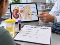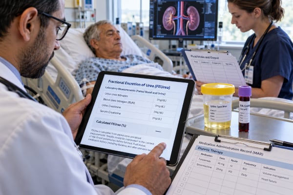
Fractional excretion of urea (FEUrea, also written FEU or FEUN) estimates the percentage of filtered urea that is excreted in urine. Clinicians mainly use it during acute kidney injury (AKI), especially when a patient has received a diuretic and the fractional excretion of sodium may be harder to interpret. Traditional teaching says an FEUrea below about 35% supports prerenal physiology, while a higher value makes intrinsic kidney injury more likely. In practice, the test has substantial overlap between causes. Sepsis, chronic kidney disease, variable protein metabolism, changing urine flow, and diuretics themselves can affect urea handling. A 2024 systematic review found only moderate diagnostic performance and did not show that FEUrea clearly outperforms FENa in patients on diuretics. FEUrea is therefore a supporting clue, not a definitive test. It is most useful when interpreted with serum creatinine trends, urine output, volume status, urinalysis, urine sediment, medications, and the clinical cause of AKI.
- FEUrea uses paired urine and blood urea and creatinine values to estimate renal urea handling.
- An FEUrea below about 35% traditionally supports a prerenal pattern, but this cutoff is not definitive.
- FEUrea is often considered after diuretics because urine sodium and FENa can rise directly from the drug effect.
- Recent evidence shows FEUrea has limited accuracy and is not consistently superior to FENa, even with diuretic use.
- AKI with very low urine output, high potassium, severe acidosis, breathlessness, or confusion needs prompt medical evaluation.
Table of Contents
- What FEUrea Measures and How It Is Calculated
- FEUrea Cutoffs and Traditional Interpretation
- Why Diuretics Matter
- Limitations and Misleading Results
- FEUrea Versus FENa and Other AKI Tests
- Sample Timing and Calculation Examples
- Follow-Up After an FEUrea Result
What FEUrea Measures and How It Is Calculated
Urea is a nitrogen-containing waste product produced mainly from protein metabolism. It is freely filtered by the glomeruli, but unlike creatinine, a substantial portion is reabsorbed along the nephron. The amount reabsorbed changes with urine flow and hormonal signals, which is why urea can provide information about the kidney’s response to low effective circulating volume.
The common formula is:
FEUrea (%) = (urine urea × serum creatinine) ÷ (serum urea × urine creatinine) × 100
Some laboratories report urea nitrogen rather than urea. The same concept applies as long as the serum and urine urea measurements use corresponding units. Urine and blood should be collected close together.
During reduced kidney perfusion, neurohormonal responses increase sodium and water reabsorption. Slower tubular flow and increased proximal reabsorption can also increase urea reabsorption. As a result, a smaller fraction of filtered urea may appear in the urine, producing a low FEUrea.
That physiologic pattern is related to the traditional BUN/creatinine ratio, which can also rise when urea reabsorption increases. However, BUN is influenced by protein intake, gastrointestinal bleeding, catabolic state, corticosteroids, liver function, and nutrition, so neither measurement is specific for volume depletion.
FEUrea describes handling of a filtered substance. It does not measure structural kidney damage directly, and it does not prove that kidney function will normalize after fluids.
FEUrea Cutoffs and Traditional Interpretation
The most commonly taught threshold is 35%.
| FEUrea result | Traditional meaning | How to use it |
|---|---|---|
| <35% | Supports prerenal physiology or avid urea reabsorption | Useful only when the clinical setting fits; not diagnostic by itself |
| 35%–50% | Overlap/indeterminate zone | Little value without other findings |
| >50% | Can support intrinsic tubular injury | Many exceptions occur |
These ranges should not be treated like a laboratory reference interval. The biologic distributions of prerenal AKI and intrinsic AKI overlap.
In a 2024 meta-analysis, an FEUrea threshold above 35% used to identify intrinsic rather than prerenal AKI had pooled sensitivity of about 66% and specificity of about 75%. That level of accuracy can shift probability, but it leaves many false-positive and false-negative results.
A low FEUrea is therefore best phrased as consistent with increased renal urea conservation, not “proof of dehydration.” Heart failure, cirrhosis, and other states with low effective arterial volume can produce similar physiology despite excess total body fluid.
A higher FEUrea can support tubular dysfunction, but it should not automatically be labeled acute tubular necrosis. The acute kidney injury blood test panel, urine output, urinalysis, and clinical course remain more fundamental.
Why Diuretics Matter
FEUrea became popular partly because loop and thiazide diuretics directly increase urinary sodium excretion. A patient can be volume depleted yet have a urine sodium and FENa that appear “too high” for classic prerenal AKI after receiving furosemide.
Because urea handling is not directly blocked at the same transport site as sodium by common loop and thiazide diuretics, FEUrea was proposed as a more reliable marker in this setting. This idea has physiologic appeal, but clinical data are less convincing than the traditional teaching suggests.
The 2024 meta-analysis comparing FEUrea with FENa found that FEUrea did not provide a clearly superior alternative. Among patients receiving diuretics, FEUrea had lower sensitivity but higher specificity than FENa for identifying intrinsic AKI in the included studies. The overall conclusion was that FEUrea has limited utility.
Timing also matters. A diuretic dose can abruptly change urine sodium, urine flow, and indirectly urea handling. If the patient has received IV fluid, vasopressors, diuretics, or other interventions, a spot urine measurement reflects the physiology after those changes.
This does not make FEUrea useless. It means the result should be interpreted as one data point rather than as a correction that “fixes” FENa.
Limitations and Misleading Results
FEUrea can be altered by several factors beyond simple kidney perfusion.
Sepsis and critical illness
Sepsis-associated AKI involves hemodynamic, inflammatory, and tubular changes that do not always follow the classic prerenal-versus-acute-tubular-necrosis model. Urea production may also increase during catabolism. A low or high FEUrea can therefore be difficult to assign to one mechanism.
Chronic kidney disease
CKD changes baseline tubular handling and reduces the reserve available to respond to acute changes. Fractional excretion measurements become less specific for a new process. The CKD blood test panel and prior creatinine values are important for separating chronic from acute dysfunction.
Protein intake and catabolism
Serum urea rises with high protein intake, tissue breakdown, fever, corticosteroid exposure, and gastrointestinal bleeding. It can be low with severe liver dysfunction or low protein intake. Although the FEUrea formula uses both serum and urine urea, changing production and tubular delivery can still complicate interpretation.
Urine flow and vasopressin
Urea recycling and reabsorption are tied to water handling and vasopressin. Very low urine flow, changes in osmolality, and antidiuretic states can influence the result independently of structural kidney injury.
Mixed kidney injury
Patients commonly have more than one process. A person can arrive with vomiting and low circulating volume, then develop sepsis-related tubular injury or receive nephrotoxic medication. In such cases, there may be no single “correct” fractional excretion pattern.
Obstruction, glomerular disease, and interstitial disease
Postrenal obstruction can show different fractional excretion values depending on timing. Glomerulonephritis and some interstitial diseases can have sodium- and urea-conserving patterns despite intrinsic kidney pathology. Urine microscopy, protein and blood measurements, and imaging can be more informative.
FEUrea Versus FENa and Other AKI Tests
FENa and FEUrea answer similar but not identical questions.
FENa tracks the fraction of filtered sodium excreted. It has the strongest evidence in oliguric AKI without CKD or diuretic exposure. A value below about 1% supports a sodium-conserving pattern.
FEUrea tracks the fraction of filtered urea excreted. A value below about 35% has traditionally supported prerenal physiology and is often used after diuretics.
Neither test directly measures tubular cell injury. In head-to-head evidence, neither consistently provides enough accuracy to replace clinical assessment.
A useful AKI evaluation may include:
- serum creatinine compared with baseline;
- urine output over hours;
- serum potassium and bicarbonate;
- urinalysis for blood, protein, glucose, and other findings;
- urine microscopy for granular casts, red cell casts, white cells, or tubular epithelial cells;
- medication and toxin review;
- blood pressure and assessment of congestion or volume loss;
- kidney ultrasound if obstruction is possible.
The presence of granular casts, for example, may strengthen evidence for tubular injury in the right clinical setting. Red blood cell casts or dysmorphic red cells point toward a very different process.
Newer biomarkers such as NGAL and KIM-1 can reflect tubular injury, but their availability and routine clinical role vary. The NGAL blood test is not a replacement for basic AKI evaluation.
Sample Timing and Calculation Examples
FEUrea usually requires a spot urine sample and a blood sample collected at roughly the same time. No special fasting is generally needed in acute care.
Whenever possible, the sample is most informative before major interventions, but testing should never delay treatment of shock, severe electrolyte abnormalities, pulmonary edema, or other emergencies.
Consider a patient with serum urea nitrogen of 40 mg/dL, urine urea nitrogen of 400 mg/dL, serum creatinine of 2.0 mg/dL, and urine creatinine of 100 mg/dL:
FEUrea = (400 × 2.0) ÷ (40 × 100) × 100 = 20%
That is a low FEUrea and supports avid urea reabsorption. If the patient has several days of vomiting, orthostatic symptoms, no edema, and improving creatinine after appropriate fluid replacement, the result fits the overall picture.
Now consider serum urea nitrogen 40 mg/dL, urine urea nitrogen 800 mg/dL, serum creatinine 2.0 mg/dL, and urine creatinine 80 mg/dL:
FEUrea = (800 × 2.0) ÷ (40 × 80) × 100 = 50%
That is more compatible with reduced urea conservation. If the patient also has muddy brown granular casts after prolonged hypotension, the evidence for acute tubular injury becomes stronger.
The same numbers should be interpreted differently if the patient has advanced CKD, sepsis, rapidly changing kidney function, or major catabolic illness.
A urine urea nitrogen test has other uses, including assessment of nitrogen balance and protein intake, but those applications usually involve timed urine collections rather than this acute fractional excretion calculation.
Follow-Up After an FEUrea Result
Clinicians treat the cause of AKI, not the FEUrea percentage.
If the result supports reduced effective circulating volume, the next step is to identify why. Gastrointestinal loss, bleeding, over-diuresis, poor intake, heart failure, cirrhosis, and systemic infection require different management. A low FEUrea does not automatically mean that giving more fluid is safe.
If intrinsic kidney injury remains likely, clinicians review nephrotoxic drugs, recent hypotension, infection, pigment injury, and urine sediment. Persistent AKI, severe electrolyte abnormalities, unclear diagnosis, or suspected glomerulonephritis may require nephrology consultation.
Urgent treatment may be needed for severe hyperkalemia, severe metabolic acidosis, pulmonary edema, uremic complications, or near-anuria. A potassium blood test and bicarbonate are therefore central in ongoing monitoring.
FEUrea is most valuable when used modestly: it can support or weaken a hypothesis about renal conservation during AKI, particularly when sodium-based measures are confounded. It should not be used to declare an AKI “prerenal” or “intrinsic” without the rest of the evidence.
Practical FEUrea examples
FEUrea is often ordered because a patient with AKI recently received a diuretic and the clinician wants another estimate of tubular reabsorption.
Suspected volume depletion after diuretics: Consider a patient with poor oral intake, orthostatic symptoms, an increased creatinine, and a recent loop diuretic dose. FENa may be elevated because the drug causes natriuresis. If FEUrea is below about 35%, that finding can support a low-perfusion state because urea reabsorption may still be increased. It does not prove that the kidneys are structurally normal, and it does not establish that giving fluid is safe in a patient who may also have heart failure.
Established tubular injury: In acute tubular injury, impaired reabsorptive function can produce an FEUrea above about 50%. A compatible story might include prolonged hypotension, sepsis, nephrotoxin exposure, persistent AKI, and granular casts. However, substantial overlap exists. Modern meta-analysis found only moderate sensitivity and specificity, so the percentage should be considered one component of the diagnosis.
Sepsis-associated AKI: Sepsis illustrates why simple categories fail. Altered microcirculation, inflammation, hemodynamic changes, vasopressor therapy, fluids, and tubular responses can occur together. Some patients with intrinsic injury retain low fractional excretion values, while others do not. Urine electrolytes in this setting can describe physiology but may not reliably identify a single cause.
Heart failure or cirrhosis: A patient can have total-body fluid excess but reduced effective arterial blood volume. The kidney may avidly reabsorb sodium and urea, creating a “prerenal” pattern even while edema is present. Treatment depends on congestion, perfusion, cardiac or liver status, and hemodynamics—not on FEUrea alone.
Why FEUrea is not simply a better FENa
The traditional rationale is biologically plausible: most urea reabsorption occurs in proximal and medullary segments and was thought to be less directly altered by loop or thiazide diuretics than sodium excretion. Yet urea handling is also affected by vasopressin, urine flow, catabolic state, protein intake, kidney function, and the severity and timing of illness.
A 2024 systematic review and meta-analysis found that FEUrea did not clearly outperform FENa for distinguishing prerenal from intrinsic AKI, including among patients receiving diuretics. This is an important update to older teaching. FEUrea can still be useful when it fits the clinical picture, but a cutoff such as 35% should not be treated as a diagnostic switch.
Questions to ask when FEUrea and the clinical picture disagree
If FEUrea appears “prerenal” but AKI persists, ask whether tubular injury has developed on top of reduced perfusion, whether obstruction has been excluded, and whether urine microscopy suggests another process. If FEUrea appears “intrinsic” despite a convincing volume-loss history, review timing of fluids, diuretics, nutrition, and the simultaneous blood and urine samples.
The trajectory of serum creatinine and urine output is often more informative than one calculation. Improvement after correction of a reversible hemodynamic problem supports a functional component, whereas persistent or worsening injury should prompt a broader evaluation. FEUrea is most valuable when it helps organize that evaluation rather than replace it.
A result should change a question, not end the evaluation
The most useful FEUrea report answers a narrow bedside question: does renal handling of urea fit with increased tubular reabsorption at this moment? If yes, clinicians can weigh that finding with perfusion, congestion, fluid losses, medication exposure, and urine sediment. If no, they can look harder for tubular injury or another renal process.
Repeated calculations are rarely helpful unless the clinical state has changed in a way that makes a new sample meaningful. Instead, follow serum creatinine, urine output, blood pressure, volume findings, and the suspected cause of AKI. A rapidly improving creatinine after correction of hemodynamics is clinically important regardless of whether the original FEUrea landed just above or below 35%. Likewise, progressive AKI with an apparently “prerenal” FEUrea deserves reassessment rather than reassurance.
This approach preserves what FEUrea does well—a quick physiologic clue—without asking the test to distinguish complex AKI syndromes more precisely than the evidence supports.
References
- Utility of fractional excretion of urea in acute kidney injury with comparison to fractional excretion of sodium: A systematic review and meta-analysis 2024 (Systematic Review)
- Fractional Excretion of Sodium and Urea are Useful Tools in the Evaluation of AKI: PRO 2023
- Fractional Excretion of Sodium and Urea are Useful Tools in the Evaluation of AKI: CON 2023
- Fractional Excretion of Sodium and Urea are Useful Tools in the Evaluation of AKI: COMMENTARY. 2023
- Diagnostic Performance of Fractional Excretion of Sodium for the Differential Diagnosis of Acute Kidney Injury: A Systematic Review and Meta-Analysis 2022 (Systematic Review)
- Use of Urine Electrolytes and Urine Osmolality in the Clinical Diagnosis of Fluid, Electrolytes, and Acid-Base Disorders 2021 (Review)
Disclaimer
FEUrea is an adjunctive AKI test and cannot determine the cause of kidney injury or whether a patient should receive fluids by itself. Acute kidney injury can cause dangerous electrolyte, acid-base, and fluid complications. Treatment should be based on the complete clinical picture and guided by a qualified clinician.