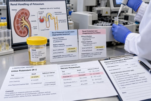
A urine potassium test measures how much potassium your kidneys are excreting. It is most useful when a blood potassium level is abnormal and the clinician needs to know whether the kidneys are responding appropriately. With low blood potassium, healthy kidneys normally conserve potassium; continued high urinary loss suggests a renal cause such as diuretics, excess aldosterone activity, or a salt-wasting tubule disorder. With high blood potassium, low urinary excretion may point toward impaired kidney excretion, reduced aldosterone effect, or low delivery of sodium and water to the potassium-secreting part of the nephron. Results can come from a 24-hour collection or a spot urine sample, and neither should be interpreted alone. Blood potassium, kidney function, acid-base status, blood pressure, medications, magnesium, and urine sodium or chloride often provide the clues that turn the urine result into a diagnosis.
- Urine potassium helps distinguish kidney potassium loss from gastrointestinal loss, poor intake, or shifts of potassium into cells.
- During hypokalemia, persistently high 24-hour urine potassium suggests inappropriate renal potassium wasting.
- A low urine potassium result during hypokalemia usually means the kidneys are conserving potassium appropriately.
- A typical adult 24-hour laboratory reference interval may be about 16–105 mmol/day, but diagnostic cutoffs depend on the blood potassium level and clinical setting.
- Severe blood potassium abnormalities, weakness, paralysis, palpitations, or ECG changes require urgent medical assessment regardless of the urine result.
Table of Contents
- What a urine potassium test measures
- Why urine potassium is ordered
- Collection methods and preparation
- How to interpret urine potassium results
- Causes of high urine potassium
- Causes of low urine potassium
- Diagnostic next steps and when results are urgent
What a urine potassium test measures
Potassium is the main positively charged ion inside cells. It is essential for normal nerve signals, muscle contraction, and heart electrical activity. The body keeps blood potassium within a narrow range by shifting potassium between cells and blood and by adjusting how much the kidneys excrete.
The kidneys perform the long-term balancing. Potassium is filtered at the glomerulus, largely reabsorbed in the earlier nephron, and then regulated in the distal nephron and collecting duct. Aldosterone, sodium delivery, urine flow, acid-base balance, dietary intake, and several kidney transport proteins influence how much potassium is secreted into urine.
A urine potassium test therefore measures kidney handling, not simply total body potassium. It becomes especially informative when interpreted against the blood potassium level. A high urine potassium value can be appropriate if blood potassium is high or intake has recently been high. The same urine value can be inappropriate if the person is hypokalemic and the kidneys should be conserving potassium.
That distinction is why a blood potassium test and urine potassium are often paired. Blood potassium answers “What is the circulating concentration now?” Urine potassium helps answer “Are the kidneys contributing to this abnormality or responding to it?”
Urine potassium may be reported as:
- 24-hour potassium excretion, usually in mmol/24 hours or mEq/24 hours. For potassium, 1 mmol equals 1 mEq.
- Spot urine potassium concentration, usually in mmol/L or mEq/L.
- A urine potassium-to-creatinine ratio, which partially corrects for how dilute or concentrated a spot sample is.
- Fractional excretion of potassium, calculated from paired blood and urine potassium and creatinine values in selected situations.
A 24-hour collection is often the clearest way to estimate daily loss, but a spot sample can be useful when a rapid answer is needed or a full-day collection is impractical.
Why urine potassium is ordered
The most common reason for urine potassium testing is unexplained hypokalemia, meaning a blood potassium level below the laboratory reference range, commonly below about 3.5 mEq/L. Hypokalemia can result from potassium leaving the body, moving into cells, or less often from inadequate intake alone. Urine testing helps separate renal loss from nonrenal causes.
For example, diarrhea can cause substantial potassium loss through the gastrointestinal tract. If the kidneys are functioning normally, they usually respond by sharply reducing potassium excretion. By contrast, a loop or thiazide diuretic can make the kidneys continue losing potassium even when the blood level is low.
Clinicians may also order urine potassium when hyperkalemia is present. In that setting, the question is whether the kidneys are excreting enough potassium for the degree of elevation. Chronic kidney disease, acute kidney injury, low aldosterone activity, medications that block the renin-angiotensin-aldosterone system, potassium-sparing diuretics, and reduced distal sodium delivery can all limit potassium excretion. A kidney function blood test panel is usually essential to interpret this pattern.
Other reasons for testing include:
- persistent low potassium despite replacement;
- suspected diuretic effect or diuretic exposure;
- high blood pressure with unexplained hypokalemia;
- metabolic alkalosis or metabolic acidosis of unclear cause;
- suspected Bartter syndrome, Gitelman syndrome, renal tubular acidosis, or another tubulopathy;
- suspected mineralocorticoid excess, such as primary aldosteronism;
- monitoring of selected kidney or endocrine disorders;
- assessment of dietary potassium exposure in research or specialized clinical settings.
Urine potassium is not a general screening test for whether someone eats “enough potassium.” A single spot concentration changes too much with hydration and timing. Repeated 24-hour collections can estimate usual intake more closely in people with stable kidney function, but clinical interpretation still requires context.
Collection methods and preparation
A 24-hour urine potassium test measures total excretion over a complete day. A major reference laboratory lists an adult interval of 16–105 mmol per 24 hours, but that is a population reference interval rather than a universal threshold for renal potassium wasting.
For a typical 24-hour collection, you discard the first morning urine, record that start time, then collect every urine specimen for the next 24 hours and include the final specimen at the same time the following morning. Follow the laboratory’s storage instructions. Missing urine makes total excretion look falsely low, while collecting for too long can make it appear falsely high.
A spot urine sample is easier. It may be collected at the same time as blood potassium and creatinine so the clinician can calculate a urine potassium-to-creatinine ratio or another renal-handling measure. Spot concentrations are strongly affected by urine volume: 25 mmol/L in very dilute urine does not mean the same thing as 25 mmol/L in a highly concentrated sample.
Preparation depends on the clinical question. Unless specifically instructed otherwise, keep your usual diet and fluid intake so the sample reflects normal conditions. Sudden heavy use of potassium-rich foods or salt substitutes can change excretion. So can vomiting, diarrhea, strenuous exercise, and a large change in fluid intake.
Medication review is crucial. Diuretics are among the most common reasons for renal potassium loss. Other relevant drugs include mineralocorticoids, glucocorticoids in some settings, certain antibiotics, amphotericin B, laxatives, insulin, beta-agonists, ACE inhibitors, angiotensin receptor blockers, mineralocorticoid receptor antagonists, and potassium supplements. Do not stop prescribed medication solely to alter the test unless the ordering clinician gives explicit instructions.
Magnesium deserves special attention. Low magnesium can make renal potassium wasting persist and can make hypokalemia difficult to correct. A magnesium blood test is therefore often part of the workup when potassium remains low.
How to interpret urine potassium results
The most important rule is that the same urine potassium number can mean different things depending on the blood potassium level. Reference intervals describe what is seen in a laboratory population; diagnostic interpretation asks whether the kidneys are responding appropriately to the current physiologic need.
During hypokalemia, the kidneys should reduce potassium excretion. Different clinical references use somewhat different cutoffs, but a 24-hour urine potassium above roughly 15–30 mEq/day during confirmed hypokalemia raises concern for renal potassium wasting. Some laboratories use more than 30 mEq/day as a clear sign that kidney loss is inappropriate. Lower values support appropriate renal conservation, especially when the collection is complete.
For a spot sample, clinicians often use a potassium-to-creatinine ratio rather than potassium concentration alone. A ratio above roughly 13 mEq potassium per gram of creatinine has been used as evidence of renal potassium loss in adults, although spot measurements have important variability and are not perfect. Units matter, so a value should never be compared with a cutoff unless the units match.
Interpretation then branches according to acid-base status and blood pressure:
| Pattern | Common diagnostic direction |
|---|---|
| Low blood K + low urine K | GI loss, low intake, or intracellular shift is more likely |
| Low blood K + high urine K + metabolic alkalosis | Diuretics, vomiting-related renal effects, Bartter/Gitelman syndromes, or mineralocorticoid excess |
| Low blood K + high urine K + metabolic acidosis | Renal tubular acidosis, ketoacidosis, or other renal potassium losses |
| Low blood K + high urine K + hypertension | Primary aldosteronism or another mineralocorticoid-like state becomes more important |
| High blood K + unexpectedly low K excretion | Reduced GFR, low aldosterone effect, medication effect, or limited distal sodium/flow may be contributing |
Urine chloride often sharpens the evaluation of metabolic alkalosis. Renin and aldosterone help evaluate hypertension with renal potassium wasting. Serum bicarbonate, chloride, magnesium, and creatinine can also be decisive.
The transtubular potassium gradient (TTKG) appears in older diagnostic algorithms, but it depends on assumptions about urine osmolality, sodium delivery, and collecting-duct physiology that often do not hold. Many contemporary clinicians prefer direct urine potassium measures, creatinine-normalized ratios, and the overall biochemical pattern instead of relying heavily on TTKG.
Causes of high urine potassium
High urine potassium does not automatically mean disease. If blood potassium is normal and dietary intake is high, greater excretion can simply reflect normal balance. The result becomes concerning when urine loss remains high despite hypokalemia.
Diuretics are a common cause. Loop and thiazide diuretics increase sodium delivery to the distal nephron and stimulate mechanisms that promote potassium secretion. They can also activate the renin-angiotensin-aldosterone system through volume loss. A recent or intermittent diuretic exposure can complicate interpretation because urine electrolytes may change over time.
Mineralocorticoid excess can cause renal potassium wasting, often with hypertension and metabolic alkalosis. Primary aldosteronism is an important example. Renin and aldosterone testing, interpreted under appropriate collection conditions and medication considerations, may be needed when this pattern is suspected.
Bartter and Gitelman syndromes are inherited salt-wasting disorders that increase renin and aldosterone and lead to renal potassium loss. Both can produce hypokalemic metabolic alkalosis with normal or low blood pressure. Gitelman syndrome commonly includes low magnesium and low urinary calcium, whereas several Bartter forms are associated with higher urinary calcium. Genetic testing may be used when clinical and biochemical findings support an inherited tubulopathy.
Renal tubular acidosis and other tubular disorders can also waste potassium. Distal and proximal renal tubular acidosis may produce hypokalemia with metabolic acidosis. Urine pH, urine ammonium surrogates, bicarbonate, chloride, and the clinical setting help distinguish these conditions.
Other causes include osmotic diuresis, some forms of kidney recovery after acute tubular injury, high-dose or chronic certain antibiotics, amphotericin B, high glucocorticoid activity, and magnesium deficiency. An acute kidney injury blood test panel can be useful when changing kidney function is part of the problem.
A high urine potassium result in a person with normal or high blood potassium may simply show that the kidneys are appropriately eliminating potassium. In hyperkalemia, the clinician is more concerned when excretion is lower than expected for the severity of the blood abnormality.
Causes of low urine potassium
During hypokalemia, low urinary potassium is usually a physiologically appropriate response. It suggests the kidneys are trying to conserve potassium, which shifts attention toward gastrointestinal losses, intracellular shifts, or low intake.
Diarrhea is a classic cause of extrarenal potassium loss. The body loses potassium-rich fluid through the intestine, and functioning kidneys respond by reducing urinary potassium. The associated acid-base pattern is often metabolic acidosis because stool also contains bicarbonate, although the exact pattern varies with the illness.
Vomiting is more complicated. Direct potassium loss in vomit is not usually the entire explanation. Volume depletion, chloride loss, alkalosis, and secondary aldosterone activation can cause the kidneys to lose potassium, so urine potassium may not always be low. Urine chloride is often particularly useful in this setting.
Intracellular shifts can lower blood potassium without a major loss from the body. Insulin, beta-adrenergic stimulation, alkalosis, and hypokalemic periodic paralysis can move potassium into cells. In a pure shift, the kidneys generally reduce excretion once they sense the lower blood level.
Low potassium intake alone rarely causes marked hypokalemia in a healthy person because the kidneys can conserve potassium efficiently, but it can contribute when combined with other losses. Poor nutrition, restrictive diets, or prolonged inadequate intake can matter more in frail or ill patients.
In hyperkalemia, low urine potassium has a different meaning. It may reflect reduced filtration or impaired distal potassium secretion. Chronic kidney disease, acute kidney injury, hypoaldosteronism, diabetes-related hyporeninemic hypoaldosteronism, ACE inhibitors, ARBs, NSAIDs, trimethoprim, and potassium-sparing diuretics are among the possibilities. A creatinine blood test and eGFR help determine whether impaired filtration is contributing.
A low spot urine potassium concentration can also simply reflect very dilute urine, which is why concentration alone is a weak basis for diagnosis.
For the same reason, a spot urine potassium result should usually be interpreted with urine creatinine or with the overall urine concentration. A value reported in mEq/L answers a different question from a 24-hour total reported in mEq/day, and the two should not be compared directly.
Diagnostic next steps and when results are urgent
If urine potassium is abnormal, the next step is usually to confirm the blood potassium value and classify the pattern rather than treating the urine number itself. Blood tests often include creatinine, eGFR, bicarbonate, chloride, magnesium, glucose, and sometimes renin and aldosterone. Urine sodium and chloride may help determine whether the distal nephron is receiving enough sodium and whether volume or chloride depletion is present.
The clinician will also review recent gastrointestinal symptoms, blood pressure, diet, supplements, licorice products, and prescription or nonprescription drugs. When the biochemical pattern points toward an inherited salt-wasting syndrome, genetic testing may be considered. When primary aldosteronism is suspected, endocrine testing follows a separate standardized pathway.
Treatment depends on the cause. Potassium replacement may correct the immediate deficit, but recurrent renal potassium loss will continue until the driver is addressed. Correcting low magnesium can be necessary for potassium to normalize. Medication changes, treatment of diarrhea or vomiting, management of an endocrine disorder, or targeted therapy for a tubulopathy may be needed.
Urine potassium itself is rarely an emergency result. The blood potassium level and symptoms determine urgency. Severe hypokalemia, especially around 2.5 mEq/L or lower, can cause weakness, paralysis, respiratory problems, and dangerous heart rhythms. Hyperkalemia can also produce muscle weakness and life-threatening conduction abnormalities. Palpitations, fainting, severe weakness, paralysis, chest symptoms, or an abnormal ECG warrant urgent assessment.
For a nonurgent result, useful questions include: Was my blood potassium low or high at the time? Was the urine a complete 24-hour collection? Does the result show appropriate kidney conservation or excessive renal loss? What do my bicarbonate, magnesium, urine chloride, blood pressure, renin, aldosterone, and kidney function show? Those details matter far more than whether the urine potassium sits just inside or outside a printed reference interval.
References
- Potassium Disorders: Hypokalemia and Hyperkalemia 2023 (Review)
- Renal Hypokalemia: An Endocrine Perspective 2024 (Review)
- Potassium homeostasis – Physiology and pharmacology in a clinical context 2023 (Review)
- Pathophysiologic approach in genetic hypokalemia: An update 2023 (Review)
- 24-Hour Urinary Sodium and Potassium Excretion and Cardiovascular Risk 2022
- Potassium, 24 Hour, Urine 2026
Disclaimer
Urine potassium should be interpreted with the blood potassium level, kidney function, acid-base status, medications, and the clinical reason for testing. This article provides general education and does not replace individualized medical care. Seek urgent assessment for severe weakness, paralysis, fainting, palpitations, or known severe blood potassium abnormalities.