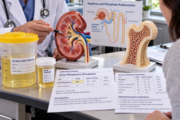
A urine phosphorus test measures how much phosphorus, mainly in the form of phosphate, your kidneys remove into urine. The result can help show whether the kidneys are conserving phosphate appropriately or losing too much, especially when a blood phosphorus level is abnormal. It may also be included in a 24-hour urine study for people with kidney stones. Because urine phosphorus changes with diet, hormones, kidney function, and the timing of collection, a high or low number is rarely meaningful by itself. The most useful interpretation compares urine phosphorus with blood phosphorus, creatinine, calcium, parathyroid hormone, vitamin D status, and the clinical reason for testing. In some cases, clinicians calculate fractional excretion of phosphate or the tubular maximum for phosphate reabsorption to determine whether a low blood phosphorus level is being caused by kidney phosphate wasting.
- A 24-hour urine phosphorus test measures total phosphate excretion over a full day; random urine phosphorus is harder to interpret by itself.
- High urine phosphorus can reflect a high-phosphate diet, parathyroid hormone effects, certain medicines, or renal phosphate wasting.
- Low urine phosphorus often means the kidneys are conserving phosphate because intake or absorption is low, but it can also occur with reduced filtration.
- When blood phosphorus is low, an inappropriately high urine phosphate loss points toward a kidney cause rather than poor intake alone.
- Reference ranges vary substantially by laboratory and diet; the result should be interpreted with the lab’s interval and related blood tests.
Table of Contents
- What the urine phosphorus test measures
- Why the test is ordered
- How the test is collected and prepared for
- Understanding urine phosphorus results
- What high urine phosphorus can mean
- What low urine phosphorus can mean
- How clinicians confirm the cause and what happens next
What the urine phosphorus test measures
Phosphorus is an essential mineral used in bone, cell membranes, energy metabolism, and acid-base chemistry. In blood and urine, most laboratory testing measures inorganic phosphate. The intestines absorb phosphate from food, bone stores or releases it as needed, and the kidneys make the final adjustment by deciding how much filtered phosphate to reabsorb and how much to excrete.
Most filtered phosphate is reclaimed in the proximal tubule, the first major segment of the kidney tubule. This reabsorption is strongly influenced by parathyroid hormone (PTH) and fibroblast growth factor 23 (FGF23). Both hormones can reduce phosphate reabsorption and increase urinary phosphate loss. Vitamin D, dietary intake, acid-base status, and kidney function also affect the system.
A urine phosphorus result therefore answers a different question from a blood phosphorus test. Blood phosphorus shows the concentration circulating at one point in time. Urine phosphorus shows what the kidneys are excreting. Looking at the two together can reveal whether the kidney response makes physiologic sense.
For example, if blood phosphorus is low, healthy kidneys should usually conserve phosphate. A urine result that remains relatively high despite hypophosphatemia suggests inappropriate renal loss. If blood phosphorus is high, increased urine excretion may be an appropriate attempt to lower the blood level, provided kidney filtration is adequate.
There are three common ways urine phosphate handling is assessed:
- 24-hour urine phosphorus: measures the total amount excreted over a full collection period and is often the most directly interpretable urine result.
- Random urine phosphorus: reports concentration in a single specimen. Hydration can change the concentration substantially, so it is commonly interpreted with urine creatinine or used in a calculated index.
- Renal phosphate handling calculations: fractional excretion of phosphate, tubular reabsorption of phosphate, and TmP/GFR combine blood and urine measurements to ask whether the kidneys are conserving or wasting phosphate appropriately.
No single method is best for every situation. The correct choice depends on whether the clinician is investigating hypophosphatemia, hyperphosphatemia, kidney stones, a tubular disorder, or a broader bone-mineral problem.
Why the test is ordered
Urine phosphorus testing is most useful when the question is not simply “Is phosphorus high or low?” but “Why is it abnormal?” It is usually ordered as part of a targeted evaluation rather than as a routine screening test.
A clinician may request urine phosphorus when a blood test shows persistent low phosphorus. The key distinction is whether phosphate is being lost through the kidneys or whether the body is receiving or absorbing too little. Kidney phosphate wasting can occur with hyperparathyroidism, FGF23-mediated disorders, Fanconi syndrome, some inherited phosphate transport disorders, and certain medicines. Measuring urine phosphate helps narrow the possibilities.
Testing can also be useful with high blood phosphorus, although chronic kidney disease is often identified primarily through blood tests because reduced filtration can limit phosphate excretion. In that setting, urine phosphorus may be interpreted alongside eGFR, creatinine, calcium, PTH, and vitamin D.
Another common setting is kidney stone evaluation. Comprehensive 24-hour urine panels may include phosphorus together with urine volume, calcium, oxalate, citrate, uric acid, sodium, potassium, magnesium, sulfate, and pH. Phosphate affects calcium-phosphate chemistry and can contribute to urinary supersaturation, but stone risk depends on the full urine profile rather than phosphorus alone.
Urine phosphorus may also be ordered when clinicians suspect:
- proximal tubular dysfunction or Fanconi syndrome;
- unexplained bone pain, fractures, rickets, or osteomalacia with low blood phosphorus;
- primary or secondary hyperparathyroidism;
- tumor-induced osteomalacia or another FGF23-mediated condition;
- a genetic hypophosphatemic disorder;
- medication-related phosphate loss;
- unusual calcium-phosphate balance in a person with recurrent stones.
Because phosphorus regulation overlaps with calcium and PTH, a PTH test often provides important context. A result that looks abnormal in isolation can become understandable once those related values are available.
How the test is collected and prepared for
The two main collection methods are a 24-hour urine collection and a random or spot sample. The 24-hour method is generally preferred when the goal is to measure total phosphorus excretion or to evaluate kidney stone chemistry.
For a 24-hour collection, the laboratory usually provides a container and specific instructions. A typical process is:
- On the starting morning, urinate into the toilet and record the time. That first specimen is not collected.
- Collect every urine specimen after that for the next 24 hours, including overnight urine.
- At the same time the next morning, collect the final urine specimen to complete the 24-hour period.
- Store the container exactly as the laboratory instructs. Some laboratories require refrigeration or a preservative, while others use different handling methods.
- Report the total collection time and, when requested, the total urine volume.
Missing even one substantial void can make a 24-hour result falsely low. Collecting for longer than instructed can make it falsely high. Many clinicians also check 24-hour urine creatinine to judge whether a collection appears reasonably complete for that individual.
Diet matters. Urine phosphate can rise after higher dietary phosphorus intake and fall when intake is lower. Phosphorus is found naturally in dairy products, meat, fish, legumes, nuts, whole grains, and many other foods. Phosphate additives in processed foods and some beverages can also contribute substantially. Unless the clinician gives different instructions, do not make an abrupt, unusual diet change just to influence the test. A representative day is often more informative than an artificially “perfect” day.
Tell the ordering clinician about medicines and supplements. Diuretics, antacids or phosphate binders, vitamin D products, phosphate supplements, and medicines that affect the proximal tubule may change interpretation. Do not stop a prescribed medicine unless the clinician specifically tells you to do so.
A random urine sample is simpler but more sensitive to hydration. A dilute sample can have a low phosphorus concentration even when total daily excretion is normal. For this reason, random urine phosphate is often paired with urine creatinine and a blood sample collected around the same time.
Understanding urine phosphorus results
There is no single universal “normal” urine phosphorus number. Reference intervals vary because laboratories use different populations, methods, age groups, and collection conditions. Diet also has a large effect.
As one example, a major reference laboratory lists an adult 24-hour urine phosphorus interval of roughly 226 to 1,797 mg per 24 hours, while another laboratory uses narrower age- and sex-specific intervals. This difference is a good reason to use the reference interval printed on your own report rather than comparing your result with a number found elsewhere.
A result is best interpreted by asking four questions:
| Question | Why it matters |
|---|---|
| What is the blood phosphorus level? | Urine phosphate can be appropriate or inappropriate depending on the blood level. |
| Was this a complete 24-hour collection or a spot sample? | Total excretion and concentration are not interchangeable. |
| What is kidney filtration like? | Reduced GFR changes the filtered phosphate load and can alter urinary excretion. |
| What are PTH, calcium, vitamin D, and related urine chemistries? | These values help identify hormonal, tubular, bone-mineral, and stone-related causes. |
When blood phosphorus is low, clinicians often shift from absolute 24-hour excretion to renal-handling calculations. Fractional excretion of phosphate (FE phosphate) estimates the percentage of filtered phosphate that appears in urine. A higher-than-expected fractional excretion during hypophosphatemia suggests the kidney is failing to conserve phosphate. Thresholds vary with the clinical method and blood phosphate level, so the calculation should be interpreted in context rather than by a single cutoff taken from a general reference.
Tubular reabsorption of phosphate (TRP) expresses the fraction reabsorbed, while TmP/GFR estimates the maximum amount of phosphate the kidney tubules can reabsorb relative to filtration. TmP/GFR is particularly useful in persistent hypophosphatemia because it accounts more directly for the relationship between filtered phosphate and tubular transport.
The central idea is simple: a urine phosphorus value can be “normal” by the laboratory range but still be physiologically inappropriate. If the blood phosphorus is very low, the kidney should usually be conserving phosphate strongly. A mid-range urine value may therefore represent excessive loss in that setting.
What high urine phosphorus can mean
High urine phosphorus is called phosphaturia. Sometimes it is an appropriate response to diet or blood phosphorus; other times it signals excessive kidney phosphate loss.
High dietary phosphorus intake is one of the most straightforward causes. A person eating large amounts of phosphate-rich foods or phosphate additives may excrete more phosphorus over 24 hours. If blood phosphorus is normal and the rest of the evaluation is reassuring, dietary intake may explain the finding.
High PTH levels can increase urine phosphate by reducing proximal tubular reabsorption. This is one reason phosphorus, calcium, PTH, and kidney function are often evaluated together. Primary hyperparathyroidism may produce low or low-normal blood phosphorus with increased renal phosphate loss. Secondary hyperparathyroidism can occur in chronic kidney disease or vitamin D deficiency, but the pattern depends on kidney function and disease stage.
FGF23 excess is another major cause of renal phosphate wasting. FGF23 lowers kidney phosphate reabsorption and suppresses active vitamin D production. It is central to several inherited hypophosphatemic disorders and tumor-induced osteomalacia. These conditions usually require specialist evaluation because diagnosis depends on the biochemical pattern, symptoms, imaging, and sometimes genetic testing.
Proximal tubule injury or Fanconi syndrome can cause phosphate to spill into urine together with other substances that are normally reabsorbed. Depending on the cause, urine glucose may be present despite normal blood glucose, and there may be bicarbonate loss, aminoaciduria, uric acid wasting, or low-molecular-weight protein loss. Certain medicines, toxins, monoclonal gammopathies, and inherited diseases can injure this part of the nephron.
Some medications can cause renal phosphate wasting directly or indirectly. Examples include particular antiviral agents, chemotherapy drugs, and treatments associated with proximal tubular dysfunction. Intravenous iron formulations can also trigger FGF23-mediated hypophosphatemia in susceptible people, sometimes with substantial phosphate loss.
In kidney stone evaluations, higher 24-hour urine phosphorus may accompany a diet pattern rich in animal protein or phosphorus additives, or it may reflect altered phosphate handling. Higher urine phosphorus has been associated with stone formation in observational research, but it is not usually treated as an isolated stone-risk target. Clinicians place more weight on the complete 24-hour profile, including urine volume, calcium, citrate, oxalate, sodium, pH, and supersaturation results.
What low urine phosphorus can mean
Low urine phosphorus usually means either that less phosphate is entering the body, the kidneys are conserving phosphate, or the filtered amount is reduced. The blood result determines which explanation is most plausible.
If blood phosphorus is low and urine phosphate is also low, the kidneys may be responding appropriately by conserving phosphate. Possible causes include low dietary intake, poor intestinal absorption, prolonged use of phosphate-binding antacids or prescribed binders, severe vitamin D deficiency, chronic diarrhea, or other malabsorption states. Redistribution of phosphate into cells can also lower blood phosphorus without producing renal wasting; examples include refeeding, insulin treatment, and respiratory alkalosis.
If kidney function is substantially reduced, total phosphate excretion may fall because less plasma is filtered. This does not necessarily mean total body phosphorus is low. In advanced chronic kidney disease, blood phosphorus may rise even though each remaining nephron is excreting a greater fraction of its filtered phosphate. This is why an isolated 24-hour urine amount should not be used as a stand-alone measure of phosphate balance in CKD. A broader kidney function blood test panel provides the filtration context needed to interpret the result.
A low random urine phosphorus concentration can also result from dilute urine. Someone who drinks a large amount of fluid may produce a low concentration simply because the urine contains more water. A 24-hour total or a creatinine-normalized measure is usually more informative.
The practical distinction is between appropriate conservation and reduced excretion for another reason. When the blood phosphorus level is low, low urinary phosphate generally argues against ongoing renal phosphate wasting. When blood phosphorus is high, low urinary phosphate can point toward impaired kidney excretion, especially if eGFR is reduced.
How clinicians confirm the cause and what happens next
An abnormal urine phosphorus result is usually a starting point, not a diagnosis. The next step is to confirm the pattern and connect it with symptoms, blood values, kidney function, diet, and medications.
Common follow-up tests include serum phosphorus, calcium, creatinine, eGFR, bicarbonate, magnesium, alkaline phosphatase, PTH, and 25-hydroxy vitamin D. When renal phosphate wasting is suspected, clinicians may calculate FE phosphate, TRP, or TmP/GFR. FGF23 testing may be appropriate when the pattern suggests an FGF23-mediated disorder, but it is not needed for every case.
A clinician may also look for evidence of generalized proximal tubule dysfunction using urinalysis and blood chemistry. If chronic kidney disease is part of the picture, phosphate interpretation should be integrated with CKD stage and the broader mineral-bone disorder assessment rather than treated as a separate number. A renal function panel can supply several of these related measurements in one set of blood tests.
For kidney stone patients, the response is usually based on the complete 24-hour urine pattern. Increasing fluid intake, reducing excessive sodium, adjusting dietary protein or phosphate additives, maintaining appropriate dietary calcium, or using targeted medication may be recommended depending on the abnormalities present. A phosphorus value alone does not determine the plan.
Seek prompt medical attention if an abnormal phosphorus result occurs with severe muscle weakness, confusion, trouble breathing, seizures, new abnormal heart rhythm symptoms, or severe illness. These symptoms are more closely related to serious disturbances in blood phosphorus or other electrolytes than to the urine result itself.
For nonurgent abnormalities, the most useful questions to ask are: Was the collection complete? What was my blood phosphorus at the same time? Is my kidney response appropriate for that blood level? Are PTH, vitamin D, or kidney function abnormal? Does the pattern suggest diet, medication, a tubular problem, or a hormonal cause? Those questions turn a raw urine number into clinically useful information.
References
- Understanding renal phosphate handling: unfinished business 2023 (Review)
- Acquired disorders of phosphaturia: Beyond tumor-induced osteomalacia 2023 (Review)
- Reflections on TRP and TP/GFR in the definition of renal phosphate loss: conceptual review 2023 (Review)
- 24-Hour Urinary Chemistries and Kidney Stone Risk 2024
- KDIGO 2024 Clinical Practice Guideline for the Evaluation and Management of Chronic Kidney Disease 2024 (Guideline)
- Phosphorus, 24 Hour, Urine 2026
Disclaimer
Urine phosphorus results should be interpreted with blood phosphorus, kidney function, diet, medications, and the reason for testing. This article is for general education and is not a substitute for diagnosis or treatment by a qualified clinician. Seek medical care promptly for severe weakness, confusion, breathing difficulty, seizures, or other serious symptoms.