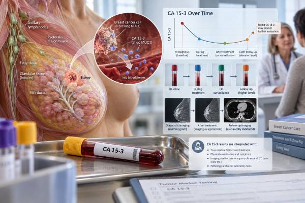
Cancer antigen 15-3, or CA 15-3, is a blood tumor marker that measures a circulating form of the MUC1 protein. Many breast cancers release increased amounts of MUC1-related antigen, especially when disease is metastatic, so CA 15-3 can be useful for following selected patients with known breast cancer. It is not accurate enough to screen healthy people for breast cancer, confirm a new breast cancer diagnosis, or routinely search for recurrence in an otherwise well person after curative treatment. A common upper reference limit is around 25–30 U/mL, but the cutoff depends on the laboratory and assay. Benign liver disease, pregnancy, inflammatory conditions, and several nonbreast cancers can also raise CA 15-3. When the marker is used in metastatic breast cancer, the most useful information usually comes from a sustained trend interpreted with symptoms, examination, and imaging. A rising CA 15-3 alone should not automatically trigger a treatment change without confirming that the overall clinical picture supports progression.
- CA 15-3 is a circulating MUC1 tumor marker used mainly in breast cancer monitoring: it is most informative in advanced or metastatic disease.
- A typical upper reference limit is about 25–30 U/mL: laboratory-specific ranges must be used.
- CA 15-3 is not recommended as a general breast cancer screening test: early breast cancers may have normal levels.
- Routine CA 15-3 surveillance after curative treatment is generally not recommended for asymptomatic patients: history, examination, and mammography remain central.
- Trends can support treatment monitoring in metastatic disease: rising or falling values should be confirmed with clinical and imaging evidence.
Table of Contents
- What CA 15-3 Measures
- Normal Range and High CA 15-3 Levels
- CA 15-3 in Breast Cancer
- Monitoring Metastatic Breast Cancer
- Recurrence and Follow-Up After Early Breast Cancer
- Noncancer Causes and Test Limitations
- What to Do With a Rising CA 15-3
What CA 15-3 Measures
CA 15-3 is not a unique molecule made only by breast cancer. It is an antigenic portion of MUC1, a large glycoprotein normally found on the surface of epithelial cells. In many cancers, MUC1 is overexpressed and abnormally glycosylated, allowing fragments to enter the circulation and be detected by immunoassays.
Breast cancer is the setting in which CA 15-3 is used most often. MUC1-related markers tend to be more frequently elevated as disease burden increases. A patient with a small localized breast cancer may have a completely normal CA 15-3, while a patient with extensive bone or liver metastases is more likely to have a high value.
This biology explains two important limitations. First, CA 15-3 lacks sensitivity for early breast cancer, so it cannot replace mammography or diagnostic imaging. Second, MUC1 is expressed by tissues other than breast tissue, so elevated values are not specific for breast cancer.
CA 15-3 is closely related to CA 27.29. Both assays measure circulating products of the MUC1 gene, but they use different antibodies and recognize somewhat different epitopes. The values are not numerically interchangeable. A person being monitored should usually stay with the same marker and laboratory rather than alternating between CA 15-3 and CA 27.29.
CEA is another serum marker sometimes followed in metastatic breast cancer. The CEA blood test measures a different glycoprotein and may provide complementary information in selected patients, but it is also nonspecific.
The major value of CA 15-3 is therefore longitudinal. If a patient’s metastatic tumor clearly produces CA 15-3, changes over time can provide an additional signal about whether disease is responding or progressing.
Normal Range and High CA 15-3 Levels
Many laboratories define normal CA 15-3 as below approximately 25 or 30 U/mL. The exact upper limit depends on the assay manufacturer, local validation, and population. The reference interval printed on the report should always take priority over a general internet cutoff.
There is no universal CA 15-3 level that diagnoses breast cancer. A result of 35 U/mL may be a mild, nonspecific elevation, while values in the hundreds can occur with advanced malignancy but still do not identify the cancer site on their own.
The result is best interpreted in one of three contexts:
No known cancer
An elevated CA 15-3 found incidentally is not a reason to diagnose breast cancer. The clinician should review breast symptoms and routine screening status, but also consider liver disease, pregnancy, autoimmune or inflammatory conditions, and other malignancies. Repeating the test may be appropriate if the elevation is modest and unexpected.
Previously treated early breast cancer
A new rise can cause understandable concern about recurrence, but routine tumor-marker surveillance in asymptomatic survivors is not generally recommended because earlier detection through serum markers has not been shown to improve outcomes enough to justify false positives and downstream testing. If a marker was nevertheless measured and is abnormal, confirmation and clinical assessment are needed before extensive imaging.
Known metastatic breast cancer
Here the test is more useful. The baseline value is compared with later values during systemic therapy. A sustained fall can support response, while a sustained rise may support progression. The marker should be used as an adjunct, not the sole measure of response.
Small differences around the upper limit may reflect normal biologic or analytical variation. Comparing 28 U/mL in one laboratory with 34 U/mL in another is particularly unreliable because assay methods differ.
CA 15-3 in Breast Cancer
CA 15-3 tends to correlate with tumor burden rather than with one specific breast cancer subtype. It can be elevated in hormone receptor-positive, HER2-positive, or triple-negative disease, although the frequency varies among studies and individual tumors.
It is more often elevated in metastatic disease than in stage I or II breast cancer. Metastases to liver and bone are commonly associated with higher values, partly because patients with those sites may have a larger total burden of MUC1-producing tumor.
CA 15-3 does not tell clinicians whether a breast lesion is benign or malignant. Diagnostic mammography, ultrasound, MRI in selected situations, and tissue biopsy are the tools that establish a new breast cancer diagnosis. Receptor testing on tumor tissue, including estrogen receptor, progesterone receptor, and HER2, guides treatment in ways CA 15-3 cannot.
The marker also does not replace genomic or molecular testing. In metastatic breast cancer, treatment decisions may depend on HER2 expression, hormone receptor status, germline or somatic BRCA alterations, PIK3CA, ESR1, PD-L1, and other biomarkers. CA 15-3 provides information about disease burden, not actionable molecular biology.
A baseline CA 15-3 can still be useful when metastatic disease is diagnosed. If it is clearly high and falls when treatment works, it becomes a convenient, low-burden companion measurement between imaging studies. If it is normal despite extensive disease, there is little reason to rely on it later because that patient’s tumor may not shed enough detectable antigen.
The blood tumor marker panel concept should not be applied to breast cancer by simply ordering many unrelated markers. Markers should be selected because they have a plausible and validated role in the known disease.
Monitoring Metastatic Breast Cancer
Metastatic breast cancer is the setting where CA 15-3 has its clearest practical role. Serial measurements can supplement clinical examination and imaging during endocrine therapy, chemotherapy, HER2-targeted therapy, antibody-drug conjugates, or other systemic treatment.
A useful monitoring sequence starts with a baseline value before or near the beginning of a new therapy. Subsequent measurements should ideally use the same laboratory and assay. The team then looks for a sustained pattern rather than reacting to one number.
Falling CA 15-3
A substantial, sustained decrease often accompanies tumor response. If pain improves, liver tests stabilize, imaging shows smaller lesions, and CA 15-3 falls, the evidence is internally consistent.
A fall does not prove every cancer site is responding. Mixed responses can occur, and some lesions may progress while others shrink. Imaging remains necessary at clinically appropriate intervals.
Stable CA 15-3
A stable marker can support stable disease when symptoms and imaging are also stable. However, a stable marker cannot rule out progression if the tumor is not a strong CA 15-3 producer.
Rising CA 15-3
A repeated upward trend can be an early warning of progression, particularly in a patient whose marker previously tracked disease reliably. It should prompt review of symptoms, examination, other laboratory results, and the timing of imaging.
An early rise after starting treatment can occasionally occur before a later decline, sometimes described as a marker flare. This is one reason treatment should not be abandoned solely because the first post-treatment CA 15-3 is higher. Clinical deterioration or clear radiographic progression carries more weight.
A marker rise is also harder to interpret during liver injury, biliary disease, or another inflammatory illness. When the trend conflicts with the rest of the clinical picture, repeating the test and looking for a noncancer explanation can prevent an unnecessary therapy change.
The goal is not to normalize CA 15-3 at all costs. The goal is to control cancer while maintaining quality of life. Some responding patients have persistently elevated markers, and some progressing patients have normal markers.
The marker can be particularly useful when disease is difficult to measure with a ruler on scans, such as diffuse bone metastases. Even there, it is only one part of assessment. Bone pain, alkaline phosphatase, functional status, and imaging findings may change on different timelines, and healing bone can look more sclerotic before it looks better radiographically. Serial CA 15-3 can add context without replacing those other measures.
Recurrence and Follow-Up After Early Breast Cancer
After surgery, radiation, and systemic therapy for stage I–III breast cancer, many patients understandably want a blood test that can provide early reassurance or warning. CA 15-3 can sometimes rise before symptoms or imaging reveal distant recurrence, but earlier biochemical detection has not established enough clinical benefit to make routine testing standard surveillance for asymptomatic patients.
The 2026 ASCO follow-up guideline update continues to emphasize risk-based clinical follow-up, with history, physical examination, and mammography as core elements. Contemporary early breast cancer guidelines similarly do not support routine serum tumor-marker testing as a replacement for evidence-based surveillance.
There are several reasons. CA 15-3 may remain normal in patients who eventually recur. It may become mildly elevated for benign reasons and lead to repeated scans, biopsies, radiation exposure, cost, and anxiety. Most importantly, finding an asymptomatic metastatic recurrence a few months earlier through a marker has not been proven to improve survival in a way that outweighs these harms.
This distinction is crucial: a marker can detect recurrence earlier without necessarily improving outcomes when used routinely. Diagnostic sensitivity and clinical usefulness are not the same thing.
If a breast cancer survivor develops new symptoms such as persistent bone pain, unexplained weight loss, shortness of breath, neurologic symptoms, abdominal swelling, or a new breast or chest-wall finding, diagnostic testing is appropriate. In that situation, clinicians may use imaging and laboratory studies, and CA 15-3 can be included if it helps answer the specific question.
For a patient at especially high recurrence risk, follow-up may be more intensive, but the strategy should be individualized by the oncology team. Emerging tools such as circulating tumor DNA are being studied for molecular recurrence, but they also require evidence that acting on an early positive result improves outcomes before they become universal surveillance standards.
Noncancer Causes and Test Limitations
CA 15-3 can be elevated without breast cancer. Reported benign associations include chronic liver disease, hepatitis, cirrhosis, benign breast conditions, some autoimmune or inflammatory diseases, and pregnancy. The degree of elevation is often mild, but overlap exists.
Other cancers can raise CA 15-3 because MUC1 is not breast-specific. Ovarian, lung, pancreatic, liver, and other epithelial malignancies may produce increased levels. This makes CA 15-3 unsuitable for identifying the origin of an unknown cancer.
Laboratory interference can also affect immunoassays. Heterophile antibodies and other uncommon analytical problems should be considered when a result is repeatedly abnormal but completely inconsistent with the patient’s clinical findings.
Assay standardization is another limitation. CA 15-3 values obtained by different commercial systems can differ because antibodies, calibrators, and epitope recognition are not identical. Longitudinal interpretation is safest with the same method.
Sensitivity is imperfect at every disease stage. Some metastatic breast cancers produce little CA 15-3. A normal value therefore cannot rule out recurrence or progression. Conversely, specificity is imperfect, so a high value cannot prove recurrence.
The marker can also lag behind or precede imaging. Neither timing pattern is inherently “wrong”; blood antigen release and structural tumor change are different biological measures. Clinicians integrate both rather than forcing them to agree at every time point.
One practical rule is to avoid making a major treatment decision from a single isolated result unless the clinical situation is urgent and other evidence supports it. Confirm the trend, ask whether the assay changed, and interpret the number in the context of the patient rather than the reference range alone.
What to Do With a Rising CA 15-3
For someone without known breast cancer, a mildly high CA 15-3 should first be confirmed and interpreted for benign causes. It is not an indication to skip directly to whole-body cancer scans. The person should remain up to date with normal breast screening and receive targeted evaluation based on symptoms and clinical risk.
For a breast cancer survivor not known to have metastatic disease, a confirmed rise should be discussed with the oncology team. The clinician will ask whether there are symptoms, examination findings, liver abnormalities, or other reasons to investigate. Imaging may be appropriate when the trend is substantial or clinically concerning, but the marker itself does not establish recurrence.
For a patient with metastatic breast cancer, compare the rise with baseline, recent imaging, symptoms, and other indicators of disease. A persistent increase that previously tracked the cancer can support progression and may prompt earlier reassessment. A small one-time rise during infection or liver inflammation is much less specific.
Do not compare a CA 15-3 value with a CA 27.29 cutoff or assume the two tests should match numerically. Although both measure MUC1-related antigen, they are different assays.
Seek prompt medical attention for new severe shortness of breath, weakness or numbness, loss of bladder or bowel control, severe headache with neurologic changes, jaundice, or rapidly worsening pain. These symptoms can signal complications of cancer or other urgent illnesses and should not wait for a tumor-marker repeat.
The most useful interpretation of CA 15-3 is therefore conditional: Is there known breast cancer? Did this patient’s tumor produce the marker before? Is the change sustained? Does the clinical and imaging picture agree? When those questions are answered, CA 15-3 can be a practical monitoring tool without being mistaken for a stand-alone cancer detector.
References
- Breast Cancer Follow-Up and Surveillance After Primary Treatment: ASCO Guideline Update 2026 (Practice Guideline)
- Advancements in non-invasive biomarkers for detection and monitoring of breast cancer recurrence 2025 (Review)
- Mucin-1: a promising pan-cancer therapeutic target 2025 (Review)
- Early breast cancer: ESMO Clinical Practice Guideline for diagnosis, treatment and follow-up 2024 (Practice Guideline)
- Breast cancer detection based on cancer antigen 15-3; emphasis on optical and electrochemical methods: A review. 2024 (Review)
- Factors influencing blood tumor marker concentrations in the absence of neoplasia 2024 (Review)
Disclaimer
CA 15-3 cannot diagnose, screen for, or exclude breast cancer by itself. It is most useful as an adjunct for selected patients with known advanced breast cancer and should be interpreted with symptoms, examination, imaging, treatment timing, and the laboratory method. A rising result should be reviewed with the treating clinician before any treatment change is made.