
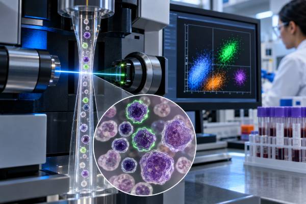
A CD22 test looks for CD22 protein on or within B cells and is used mainly to help classify B-cell leukemias and lymphomas. CD22 is a B-lineage marker that appears during B-cell development and is present on many mature B cells, while plasma cells usually lose it. In acute B-lymphoblastic leukemia (B-ALL), chronic B-cell disorders, and several B-cell lymphomas, CD22 may help confirm lineage and can also matter when a CD22-directed treatment is being considered. A “CD22-positive” result does not mean that a person has cancer. Normal B cells express CD22, and malignant B cells can show strong, weak, partial, or occasionally absent expression. The meaning depends on which cells were tested, how the test was performed, the percentage and intensity of positive cells, and the rest of the immunophenotype. Pathologists therefore interpret CD22 together with morphology, flow cytometry markers such as CD19 and CD20, genetic findings, and the clinical picture rather than as an isolated cancer test.
- CD22 is primarily a B-cell lineage marker. Positivity supports B-cell differentiation but does not by itself prove leukemia or lymphoma.
- B-ALL commonly expresses CD22, sometimes with variable or dim surface staining. Cytoplasmic CD22 can help identify early B-lineage blasts.
- There is no universal normal or “high” CD22 range. Reports usually describe the percentage of positive cells, staining intensity, and whether expression is uniform or heterogeneous.
- CD22 expression can affect treatment eligibility in some B-cell cancers. It is the target of drugs such as inotuzumab ozogamicin in B-ALL.
- Results must be read as part of a marker panel. CD19, CD20, CD10, TdT, CD34, light chains, and other markers often provide the context needed for diagnosis.
Table of Contents
- What CD22 Is and Why It Is Tested
- How CD22 Testing Is Performed
- What a CD22-Positive Result Means
- CD22 in B-Cell Acute Lymphoblastic Leukemia
- CD22 in Lymphoma and Other B-Cell Cancers
- Treatment and MRD Implications
- Limitations and Next Steps
What CD22 Is and Why It Is Tested
CD22 is a cell-surface protein that identifies B-lineage cells and helps regulate B-cell receptor signaling. Its formal name is sialic acid-binding immunoglobulin-like lectin 2, or SIGLEC-2. It is mainly expressed in the B-cell compartment and becomes detectable during B-cell maturation.
That biology makes CD22 useful in hematopathology. When a blood, bone marrow, lymph node, or other tissue sample contains an abnormal population of lymphoid cells, the laboratory needs to determine what lineage those cells belong to. No single marker is sufficient in every case, so CD22 is combined with other antigens that define B-cell, T-cell, myeloid, plasma-cell, or precursor differentiation.
Common reasons for testing CD22 include:
- classifying acute leukemia when B-ALL is in the differential diagnosis;
- characterizing a mature B-cell lymphoma or leukemia;
- confirming B-lineage differentiation when other markers are weak or altered;
- documenting antigen expression before CD22-directed therapy;
- evaluating whether CD22 expression persists after treatment; and
- helping build a leukemia-associated immunophenotype for follow-up.
The related CD19 test is also widely used to identify B-lineage cells. CD19 and CD22 overlap, but they are not redundant. A malignant clone may retain one marker while losing or reducing another, especially after targeted therapy.
CD22 is also biologically attractive as a drug target because it undergoes internalization after an antibody binds to it. That property allows an antibody-drug conjugate to carry a toxic payload into CD22-positive leukemic cells. This therapeutic role is separate from its diagnostic role, but it is one reason CD22 testing may have direct clinical relevance.
How CD22 differs from CD19 and CD20
CD19, CD20, and CD22 are often grouped together as B-cell markers, but they appear at different stages of B-cell development and behave differently in disease. CD19 is expressed relatively early and remains useful across many precursor and mature B-cell neoplasms. CD20 generally appears later, is usually absent or weak on the most immature B-ALL blasts, and is strongly associated with many mature B-cell lymphomas. CD22 can be detected in the cytoplasm of early B-lineage cells before robust surface expression develops.
This difference can resolve apparently conflicting reports. A B-ALL sample may be CD19-positive, cytoplasmic-CD22-positive, surface-CD22-dim, and CD20-negative without being contradictory. A mature lymphoma may express all three markers. After targeted therapy, one antigen can be reduced or lost while the others remain detectable. Pathologists therefore read the markers as a developmental pattern. The combination also improves confidence that a small abnormal population is truly B-lineage and helps avoid overinterpreting a single weak stain.
How CD22 Testing Is Performed
CD22 is most often measured by flow cytometry, although immunohistochemistry and other antibody-based methods can also detect it in tissue. The choice depends on the specimen and the disease being investigated.
In flow cytometry, cells from blood, bone marrow, lymph node, or another fresh specimen are mixed with fluorescent antibodies. The instrument evaluates thousands to millions of individual cells and records whether each cell expresses CD22 along with many other markers. This provides information about the percentage of positive cells, relative antigen intensity, and whether expression is uniform or restricted to a subpopulation.
For acute leukemia, laboratories may evaluate both surface CD22 and cytoplasmic CD22. Early B-cell precursors can contain CD22 inside the cell before they display as much of it on the surface. Cytoplasmic CD22 can therefore strengthen evidence of B-lineage differentiation when a blast population has an immature phenotype.
Tissue biopsies can be evaluated by immunohistochemistry, where an antibody highlights CD22-positive cells in their architectural setting. Flow cytometry often provides richer quantitative immunophenotyping, while tissue staining preserves spatial information. The best method depends on specimen availability and the diagnostic question.
A typical B-lineage panel may include CD19, CD20, CD22, CD79a, PAX5, CD10, TdT, CD34, CD45, and immunoglobulin light chains, with additional markers chosen for the suspected disease. A leukemia flow cytometry panel is designed to interpret these markers as a pattern rather than as isolated positive or negative results.
No special fasting or medication preparation is usually required for the marker itself. Preparation depends on how the sample is obtained. A blood draw is routine; a bone marrow biopsy requires local procedural preparation; a lymph node biopsy follows its own surgical or radiologic plan.
What a CD22-Positive Result Means
CD22 positivity means that the tested cells express CD22 and therefore show evidence of B-cell differentiation. It does not distinguish normal B cells from malignant B cells on its own.
A flow report may describe CD22 as positive, negative, dim, bright, partial, heterogeneous, or present on a stated percentage of abnormal cells. The exact threshold for calling a population positive depends on the assay, antibody clone, instrument, laboratory validation, disease, and intended use. That is why one laboratory’s percentage cutoff should not automatically be applied to a report from another laboratory.
| Report pattern | General meaning | Why context matters |
|---|---|---|
| CD22 positive on a B-cell population | Supports B-lineage differentiation | Normal B cells are also CD22-positive |
| Dim or partial CD22 on blasts | Can occur in B-ALL | Other precursor B-cell markers are needed for classification |
| Bright, relatively uniform CD22 | Shows substantial antigen expression | Does not identify a specific lymphoma by itself |
| Heterogeneous CD22 | Different cells within the clone show different antigen levels | May affect detection or targeted-therapy interpretation |
| CD22 absent on an abnormal B-cell population | Possible antigen loss or naturally negative/weak phenotype | B-cell lineage may still be established by other markers |
For lymphoma diagnosis, a CD22-positive population is generally evaluated with markers that define cell maturity, clonality, and subtype. For example, CD5, CD10, CD23, FMC7, CD103, CD200, cyclin D1, BCL6, and other markers may be selected depending on the suspected lymphoma. The lymphoma flow cytometry panel helps determine whether the cells are clonal and how their phenotype fits recognized disease entities.
A common misunderstanding is to treat “positive staining” as equivalent to an elevated blood tumor marker. CD22 is usually a cell-expression result, not a serum concentration. The clinical question is which cells carry CD22 and what those cells represent.
CD22 in B-Cell Acute Lymphoblastic Leukemia
Most B-ALL cases express CD22, making it useful for lineage assignment and, in selected patients, for targeted treatment. B-ALL is a malignancy of immature B-lineage lymphoblasts. Its diagnosis combines morphology, immunophenotyping, and genetic testing because modern disease classification is increasingly defined by recurrent genomic abnormalities.
A typical B-ALL blast population may express CD19, cytoplasmic CD79a, CD22, CD10, TdT, and often CD34, although the exact pattern varies. Surface immunoglobulin is usually absent in precursor B-ALL. Some subtypes show distinctive immunophenotypes that can suggest—but not prove—a particular genetic lesion.
CD22 expression in B-ALL can be heterogeneous. One patient’s blasts may show strong surface expression, while another’s are dim or only partially positive. Cytoplasmic CD22 can be especially valuable in very immature cases because it may be detectable when surface expression is limited.
CD22 should not be used alone to distinguish B-ALL from normal precursor B cells called hematogones. Hematogones can appear in normal or regenerating marrow and express a coordinated maturation pattern. Leukemic blasts usually show a more uniform, arrested, or aberrant phenotype and must be interpreted with morphology and genetics.
For patients receiving therapy, the baseline immunophenotype can also help later comparisons. However, targeted treatment can change antigen expression, so a follow-up sample should never be searched only for the exact marker pattern present at diagnosis. Modern MRD laboratories use multidimensional approaches that can recognize phenotypic shifts.
If the diagnosis is established, molecular and cytogenetic findings often carry more direct risk and treatment implications than the amount of CD22 itself. CD22 is important, but it is one part of a much larger classification system.
CD22 in Lymphoma and Other B-Cell Cancers
Many mature B-cell lymphomas and leukemias express CD22, but the marker rarely identifies a specific subtype by itself. Expression can be found across several disorders, including some diffuse large B-cell lymphomas, follicular lymphomas, marginal zone lymphomas, hairy cell leukemia, mantle cell lymphoma, and other mature B-cell neoplasms.
Because the distribution is broad, the diagnostic value comes from the combination of markers. A clonal mature B-cell population is usually established by a restricted kappa/lambda light-chain pattern or another evidence of clonality, followed by subtype-oriented immunophenotyping and genetic or tissue studies.
CD22 can be useful when another B-cell marker is weak or absent. For example, previous anti-CD20 therapy can reduce CD20 expression, making alternative B-lineage markers important. The related CD20 test remains central in many mature B-cell lymphomas, but CD20 loss does not necessarily mean the cells have stopped being B-lineage cells.
In tissue, morphology remains essential. Large atypical cells, small lymphocytes, plasmacytoid cells, nodular architecture, diffuse architecture, and the relationship to surrounding reactive cells all contribute information that an isolated flow percentage cannot provide.
CD22 expression also varies between disease sites and over time. A lymph node sample and a blood sample may contain different proportions of the malignant clone. Treatment may select for subclones with lower antigen density. For this reason, a historic “CD22-positive” result may not be sufficient when a current treatment decision depends on present antigen expression.
The practical takeaway is that CD22 is best viewed as a B-cell identity marker with therapeutic relevance, not as a lymphoma subtype label.
Treatment and MRD Implications
CD22 can be both a diagnostic marker and a treatment target, particularly in B-ALL. Inotuzumab ozogamicin is an antibody-drug conjugate that binds CD22, is internalized by the cell, and delivers calicheamicin. Its use has made careful understanding of CD22 expression clinically important in relapsed or refractory B-ALL and in some modern combination strategies.
The percentage of CD22-positive blasts and the amount of antigen per cell may influence biological sensitivity, but treatment response is not determined by one cutoff alone. Other factors include disease burden, prior therapy, genetic risk, drug exposure, cell-intrinsic resistance, and whether CD22-negative or low-expressing subclones are present.
After CD22-directed therapy, several changes can complicate testing:
- surface CD22 may decrease;
- a previously small CD22-low population may become more prominent;
- the malignant clone may show broader immunophenotypic drift; and
- assay sensitivity may change if the follow-up strategy relies too heavily on CD22.
That is why MRD assessment uses multiple markers or molecular targets. For B-ALL, minimal residual disease testing may use flow cytometry, PCR-based methods, or next-generation sequencing depending on the patient’s disease and the laboratory.
CD22 is also being studied as a target for CAR-T cells, bispecific approaches, and other antibody-based therapies, including dual-targeting strategies designed to reduce antigen escape. These approaches do not make every CD22-positive B-cell cancer an automatic candidate for CD22-directed therapy. Treatment indications depend on the exact diagnosis, regulatory approvals, treatment line, age, prior therapies, and clinical-trial availability.
For a patient reading a report, the most useful question is: “Does this CD22 result change diagnosis, measurable disease tracking, or a specific treatment option?” The hematology team can answer that only after integrating the rest of the case.
Limitations and Next Steps
CD22 testing is informative but has technical and biological limitations, so an unexpected result often needs correlation rather than immediate conclusions. Sample quality is one of the most important issues. A hemodiluted marrow aspirate can underrepresent blasts. A small tissue biopsy may miss a heterogeneous tumor population. Delays in processing can affect fragile cells and some surface antigens.
Other limitations include differences in antibody clones, fluorochromes, gating strategies, intensity scales, and positivity thresholds. A percentage reported by one laboratory is not necessarily directly comparable with a value from another laboratory. This matters especially when reviewing older reports or tracking antigen density over time.
If CD22 is unexpectedly negative in suspected B-ALL, the laboratory may rely more heavily on other B-lineage markers and cytoplasmic antigens. If CD22 is positive in a tissue lesion, the pathologist still needs to prove that the positive cells are the abnormal population rather than background B cells.
After a positive or abnormal CD22 result, common next steps can include:
- confirming the complete immunophenotype and clonality;
- reviewing marrow or tissue morphology;
- performing cytogenetic, FISH, or molecular studies appropriate to the suspected diagnosis;
- staging the disease if a lymphoma or leukemia is confirmed;
- determining whether CD22 expression has treatment implications; and
- choosing an MRD strategy if ongoing disease monitoring is needed.
A CD22 result rarely requires urgent action by itself. Urgency depends on the disease and the patient’s condition. Acute leukemia can require rapid specialist management because severe cytopenias, infection, bleeding, leukostasis, or tumor lysis risk can become emergencies. In lymphoma, rapidly enlarging masses, breathing or swallowing difficulty, neurologic deficits, or severe systemic illness also warrant prompt evaluation.
The report is therefore best interpreted with a hematologist or hematopathologist who can explain what population was tested, how convincing the B-lineage assignment is, and whether the finding changes the next diagnostic or treatment decision.
References
- CD22 as a Target for Hematological Malignancies and Autoimmune Diseases 2026 (Review)
- The Flow Cytometric Evaluation of B- and T-Lymphoblastic Leukemia/Lymphoma 2025 (Review)
- Inotuzumab ozogamicin for the treatment of adult acute lymphoblastic leukemia: past progress, current research and future directions 2024 (Review)
- Targeting CD22 for B-cell hematologic malignancies 2023 (Review)
- Efficacy and safety of CD22-specific and CD19/CD22-bispecific CAR-T cell therapy in patients with hematologic malignancies: A systematic review and meta-analysis 2022 (Systematic Review)
Disclaimer
This article provides general educational information and is not a diagnosis or treatment recommendation. CD22 results must be interpreted with the full immunophenotype, morphology, genetics, treatment history, and clinical findings by qualified hematology and pathology professionals. Seek urgent care for severe bleeding, fever with marked immune suppression, breathing difficulty, confusion, or other rapidly worsening symptoms.





