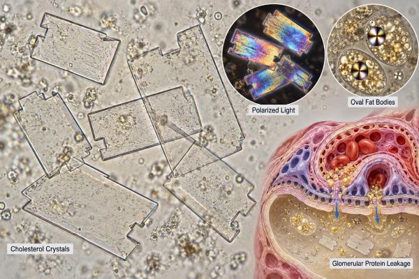
Cholesterol crystals in urine are an uncommon form of lipiduria and are most important when they appear with heavy protein loss from the kidneys. They can occur in nephrotic syndrome, where damage to the glomerular filtration barrier allows large amounts of protein and lipoprotein material to enter the urine. Under the microscope, cholesterol may appear as clear, flat plates with notched corners, often alongside oval fat bodies or fatty casts. The finding does not mean that blood cholesterol crystals are “spilling out” in a simple one-to-one way, and it does not identify the kidney disease causing the protein leak. Instead, it is a clue that should be interpreted with urine protein measurements, blood albumin, kidney function, edema, and the overall sediment. When cholesterol crystals accompany marked proteinuria, the priority is to confirm the amount of protein loss and determine whether a nephrotic pattern or another glomerular disease is present.
- Cholesterol crystals are a form of urinary lipid and are most concerning when found with heavy proteinuria.
- They often appear as large, clear rectangular plates with characteristic notched corners.
- Lipiduria in nephrotic syndrome may also include oval fat bodies and fatty casts that show a Maltese-cross pattern under polarized light.
- Cholesterol crystals do not diagnose a specific kidney disease; urine protein, serum albumin, kidney function, and clinical findings determine significance.
- New severe swelling, shortness of breath, reduced urine output, or symptoms of a blood clot need prompt medical evaluation.
Table of Contents
- What Cholesterol Crystals in Urine Mean
- How Cholesterol Crystals Look Under the Microscope
- Why Nephrotic Syndrome Causes Lipiduria
- Heavy Proteinuria and Conditions Linked to Cholesterol Crystals
- Tests That Clarify the Finding
- Treatment and Monitoring Depend on the Cause
- When Cholesterol Crystals Need Prompt Follow-Up
What Cholesterol Crystals in Urine Mean
Cholesterol crystals usually indicate lipiduria—fat-containing material in the urine—and are most clinically meaningful when there is substantial protein leakage from the kidney. They are not a routine finding in healthy urine and should prompt attention to the rest of the urinalysis rather than being interpreted alone.
The kidneys normally keep most albumin and larger lipoprotein particles in the bloodstream. When the glomerular filtration barrier becomes abnormally permeable, proteins and lipid-associated particles can cross into the filtrate. Tubular cells may take up this lipid material, and some is later seen in urine as droplets, lipid-laden cells, casts, or free crystals.
A cholesterol crystal is therefore a marker of the urinary environment, not a diagnosis by itself. The same microscopy report may have very different implications in two people. A patient with cholesterol crystals, 4+ protein on dipstick, marked edema, and low serum albumin has a very different pattern from a patient whose specimen was old, contaminated, or otherwise difficult to interpret.
The broader urine microscopy pattern in kidney disease matters. Red blood cell casts may suggest inflammatory glomerular injury, while fatty casts and oval fat bodies point toward lipid-rich proteinuria. White blood cells, bacteria, or abundant epithelial cells may indicate a separate or additional process.
Cholesterol crystals are also not a direct measure of blood cholesterol. Nephrotic syndrome commonly causes hyperlipidemia, but a urinary crystal count cannot substitute for a serum lipid panel. It simply adds another clue that the filtration barrier and tubular handling of filtered lipids may be abnormal.
How Cholesterol Crystals Look Under the Microscope
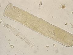
Classic cholesterol crystals are large, colorless, flat plates with one or more notched corners. They may look rectangular or square and can overlap in layers. Their sharp geometric appearance often makes them distinctive, but laboratory interpretation still depends on specimen quality and experience.
Cholesterol is strongly birefringent, meaning it interacts with polarized light in a recognizable way. Polarization can help distinguish lipid material from ordinary debris. However, the well-known “Maltese cross” appearance in nephrotic urine is more often associated with lipid droplets inside oval fat bodies or fatty casts, especially cholesterol esters and other birefringent lipids, rather than with a single free cholesterol plate.
Several lipid-related findings may appear together:
- Free cholesterol crystals: Flat, notched plates in the urine sediment.
- Oval fat bodies: Renal tubular epithelial cells or macrophage-like cells containing many lipid droplets.
- Fatty casts: Casts formed in renal tubules that contain lipid droplets or lipid-laden cells.
- Free lipid droplets: Refractile droplets that may require polarization or lipid staining for confident identification.
The presence of fatty casts in urine supports a renal source because casts form within kidney tubules. Free lipid droplets are less specific and can be confused with contaminants such as lubricants, oils, or specimen debris.
Specimen handling matters. Delayed analysis can change the appearance of cells and crystals, and contamination can introduce substances that mimic urinary lipids. A fresh, properly collected sample gives the laboratory the best chance to identify true lipiduria.
When morphology is uncertain, the laboratory should avoid overcalling a rare crystal based on one shape alone. The strength of the result rises when classic microscopy is accompanied by heavy proteinuria, hypoalbuminemia, edema, and other nephrotic features.
Why Nephrotic Syndrome Causes Lipiduria
Nephrotic syndrome creates lipiduria because severe glomerular protein leakage changes both plasma lipid metabolism and what reaches the renal tubules. The syndrome is classically defined by heavy proteinuria, low serum albumin, edema, and often hyperlipidemia.
In adults, “nephrotic-range” proteinuria has traditionally meant more than about 3.5 g of protein per day. Many clinicians now use spot urine protein-to-creatinine measurements as a practical estimate, but the exact threshold and preferred measurement can vary with the clinical setting. Importantly, nephrotic-range proteinuria is not automatically the same as full nephrotic syndrome; the blood albumin level and clinical features matter too.
When large amounts of protein and lipoprotein-associated material pass through the glomerular barrier, proximal tubular cells reabsorb some of it. These cells can accumulate lipid droplets and later appear in urine as oval fat bodies. Lipid-rich casts can form when this material becomes incorporated into the protein matrix produced in the tubules. Free cholesterol crystals can appear as lipids precipitate in the sediment.
At the same time, the liver increases lipoprotein production in response to changes associated with severe protein loss, while lipid clearance may also be altered. This contributes to the hyperlipidemia commonly seen in nephrotic syndrome. The older physiologic literature on nephrotic syndrome also documented lipiduria as a consistent feature in affected patients.
A nephrotic syndrome urine test panel is more useful than focusing on the crystal alone. Protein quantity, albumin loss, sediment findings, and kidney function help determine whether the pattern is truly nephrotic.
Lipiduria can be striking, but its absence does not rule out nephrotic syndrome. Microscopy is sample-dependent, and not every specimen contains visible cholesterol crystals or fatty casts. Likewise, visible lipid does not reveal which glomerular disease caused the barrier injury.
Heavy Proteinuria and Conditions Linked to Cholesterol Crystals
The main clinical link is severe glomerular proteinuria, not one specific kidney disease. Several primary and secondary disorders can produce nephrotic-range protein loss and therefore create a setting in which cholesterol crystals, oval fat bodies, or fatty casts appear.
Common or important causes include:
- Minimal change disease: A common cause of nephrotic syndrome in children and also seen in adults.
- Focal segmental glomerulosclerosis (FSGS): A pattern of glomerular scarring that may be primary or secondary to other conditions.
- Membranous nephropathy: An important cause of adult nephrotic syndrome, sometimes associated with autoantibodies, medications, infections, malignancy, or other disorders.
- Diabetic kidney disease: Long-standing diabetes can cause progressively heavy albuminuria and, in advanced cases, nephrotic-range protein loss.
- Amyloidosis: Abnormal protein deposits can damage glomeruli and produce marked proteinuria.
- Lupus nephritis and other immune-mediated diseases: These may produce nephrotic protein loss, hematuria, inflammatory sediment, or a mixed picture.
The amount of protein should be confirmed rather than inferred from crystals. A dipstick is a useful screen but is affected by urine concentration and mainly detects albumin. A urine protein-to-creatinine ratio estimates total protein excretion from a spot sample, while a 24-hour collection may be used when exact quantification is important.
Albumin-specific testing can also matter. The urine albumin-to-creatinine ratio is widely used to detect and stage albuminuria, especially in diabetes and chronic kidney disease. Very heavy proteinuria may require broader quantification because non-albumin proteins can contribute.
The word “cholesterol” can mislead people into assuming diet is the immediate cause of the crystal. High dietary cholesterol is not the usual explanation. The central problem is abnormal renal filtration and handling of protein and lipoprotein material. Diet may influence cardiovascular risk and serum lipids, but it does not replace evaluation of the kidney lesion causing protein loss.
Tests That Clarify the Finding
When cholesterol crystals are reported, the next step is to confirm whether there is clinically important protein loss and assess kidney function. Testing should answer whether the finding is part of nephrotic syndrome, another glomerular disease, or a less significant sediment abnormality.
Useful tests often include:
- Repeat urinalysis with fresh microscopy. This confirms the crystal and checks for protein, blood, cells, and casts. A clean, promptly analyzed specimen reduces artifacts.
- Quantitative urine protein testing. A spot protein-to-creatinine ratio or timed collection can establish whether protein loss is in the nephrotic range.
- Urine albumin measurement. Albumin quantification helps characterize glomerular leakage and provides a baseline for monitoring.
- Serum albumin and total protein. A low albumin level supports a nephrotic pattern when heavy proteinuria is present.
- Serum creatinine and estimated GFR. These show how well the kidneys are filtering and whether acute or chronic kidney dysfunction accompanies the protein loss.
- Serum lipid panel. Total cholesterol, LDL cholesterol, triglycerides, and HDL cholesterol help document the hyperlipidemia that often accompanies nephrotic syndrome.
- Cause-directed blood tests. Depending on age and presentation, clinicians may test for diabetes, autoimmune disease, infections, monoclonal proteins, or antibodies linked to membranous nephropathy.
- Kidney biopsy when indicated. Biopsy can identify the underlying glomerular lesion when the diagnosis is uncertain or when tissue results will change treatment.
A low blood albumin level is especially important when interpreted with heavy urine protein. Low albumin can also occur with liver disease, inflammation, malnutrition, or protein loss from other sites, so urine data are essential.
Microscopy should not be used as a substitute for quantitative protein testing. The number of crystals in one field does not tell how many grams of protein are lost per day, and a person can have severe nephrotic syndrome without visible cholesterol crystals in every sample.
Serial measurements are useful after treatment. Falling proteinuria and rising serum albumin generally provide a clearer measure of response than whether rare crystals are still seen on one follow-up slide.
The urine sediment can also help decide whether the process looks purely nephrotic or mixed. Heavy protein with lipiduria and a relatively bland sediment can fit a classic nephrotic pattern, whereas numerous dysmorphic red cells or red blood cell casts suggest added glomerular inflammation. That distinction can change the urgency and type of diagnostic workup, even when the amount of protein is similar.
Treatment and Monitoring Depend on the Cause
There is no treatment aimed specifically at dissolving urinary cholesterol crystals. Treatment targets the glomerular disease, the degree of proteinuria, edema, blood pressure, thrombotic risk, infection risk, and cardiovascular complications.
For many proteinuric kidney diseases, clinicians use measures that reduce intraglomerular pressure and urine protein, such as renin-angiotensin system blockers when appropriate. Some patients benefit from SGLT2 inhibitors depending on the cause, kidney function, diabetes status, and current guidelines. Primary immune-mediated diseases may require corticosteroids, rituximab, calcineurin inhibitors, or other immunosuppressive treatments, but the choice depends on the specific diagnosis rather than the presence of lipiduria.
Edema may require sodium restriction and diuretics. Severe edema can be difficult to treat because low serum albumin changes fluid distribution and diuretic response. Management is individualized to blood pressure, kidney function, body weight, and symptoms.
Hyperlipidemia is also assessed because nephrotic syndrome can markedly raise cholesterol and triglycerides. Cardiovascular risk, age, expected duration of nephrosis, and other factors influence whether lipid-lowering medication is used. The urinary crystal itself is not a target for cholesterol-lowering therapy.
Monitoring usually focuses on protein excretion, serum albumin, creatinine or eGFR, blood pressure, edema, weight, and disease-specific markers. A nephrotic-range proteinuria test can be repeated to show whether protein loss is improving.
Because nephrotic syndrome increases the risk of blood clots and infection, treatment plans sometimes include additional prevention based on individual risk. Decisions about anticoagulation are complex and should not be made from a urine microscopy result alone.
As the underlying nephrotic process improves, lipiduria often decreases. That makes cholesterol crystals useful as part of the sediment story, but quantitative protein and clinical recovery remain more important measures of progress.
Proteinuria should be followed with the same quantitative method when possible so trends are meaningful. Day-to-day urine concentration can make dipstick grades fluctuate even when total protein loss has not changed much. A consistent protein-to-creatinine or albumin-to-creatinine approach, interpreted with serum albumin and clinical edema, gives a more reliable view of response than counting cholesterol crystals on repeat microscopy.
When Cholesterol Crystals Need Prompt Follow-Up
A report of cholesterol crystals deserves timely follow-up when it comes with heavy protein, swelling, low serum albumin, blood in the urine, or reduced kidney function. The urgency depends on symptoms and the overall kidney picture.
Arrange clinical review soon if a urine report shows marked protein together with cholesterol crystals or fatty casts, especially if there is new ankle swelling, puffiness around the eyes, rapid weight gain from fluid, foamy urine, or high blood pressure. These findings may indicate substantial glomerular protein loss that needs quantification and a cause-focused evaluation.
Seek urgent care for:
- Shortness of breath, chest pain, or severe swelling that suggests fluid overload.
- One-sided leg swelling, sudden chest pain, coughing blood, or unexplained breathlessness, which can be symptoms of a blood clot.
- Fever or serious infection symptoms in a person with active nephrotic syndrome or immunosuppressive treatment.
- Markedly reduced urine output, rapidly worsening kidney function, confusion, or severe weakness.
- Severe headache, neurologic symptoms, or very high blood pressure with symptoms.
A crystal finding by itself is rarely an emergency. The risk comes from the kidney disease and its complications, not from the microscopic plate of cholesterol.
If the result is unexpected and there are no symptoms, asking whether the sample was fresh and whether the laboratory also saw protein, fatty casts, or oval fat bodies is reasonable. Repeat testing can confirm the sediment, but quantitative protein measurement should not be delayed if the initial urinalysis already shows heavy protein.
The practical interpretation is straightforward: cholesterol crystals are a clue to lipid-rich urine, and heavy proteinuria determines their importance. Confirm the protein loss, check serum albumin and kidney function, and investigate the underlying glomerular disorder rather than treating the microscopy finding in isolation.
References
- An updated approach to the evaluation of the urinary sediment 2025 (Review)
- Diagnostic Utility of Urine Microscopy in Kidney Diseases 2024 (Review)
- Nephrotic Syndrome: A Review 2024 (Review)
- Renal and Urinary Conditions: Nephrotic Syndrome 2024 (Review)
- Abnormalities of lipoprotein metabolism in patients with the nephrotic syndrome 1990
Disclaimer
This article is for general education and cannot determine why cholesterol crystals or protein are present in an individual urine sample. Heavy proteinuria, low blood albumin, swelling, or impaired kidney function should be evaluated by a clinician because nephrotic syndrome has several possible causes and complications. Seek urgent care for breathing difficulty, severe swelling, signs of a blood clot, or sharply reduced urine output.