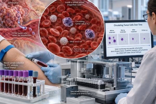
A circulating tumor cell (CTC) blood test looks for intact cancer cells that have detached from a tumor and entered the bloodstream. These cells are rare—sometimes only a few are found among billions of normal blood cells—but their presence can provide information about metastatic spread, prognosis, and treatment response in selected cancers. CTC testing is best established in metastatic breast, colorectal, and prostate cancer, where the CellSearch system has regulatory clearance as an aid to monitoring and prognosis. A high CTC count does not by itself prove that treatment has failed, and a low or undetectable count does not guarantee that cancer is absent. Different CTC technologies capture and define cells in different ways, so results are not interchangeable across platforms. CTC testing is also different from circulating tumor DNA (ctDNA): CTC assays analyze whole tumor cells, while ctDNA assays examine DNA fragments shed into blood. In routine care, CTC results must be interpreted with imaging, symptoms, pathology, and other biomarkers.
- CTC tests search for intact tumor cells in blood; they are a type of liquid biopsy but are different from ctDNA testing.
- For CellSearch, historically validated unfavorable counts are ≥5 CTCs per 7.5 mL in metastatic breast or prostate cancer and ≥3 per 7.5 mL in metastatic colorectal cancer.
- Higher CTC counts are generally associated with shorter progression-free and overall survival in validated metastatic-cancer settings.
- A falling CTC count during treatment can be reassuring, but treatment decisions should not rely on the CTC number alone.
- CTC testing is not an established general cancer-screening test, and many early cancers release too few detectable cells.
Table of Contents
- What Circulating Tumor Cells Are
- How CTC Blood Testing Works
- CTC Counts and What High Levels Mean
- How CTCs Are Used for Cancer Monitoring
- CTC Testing Compared With ctDNA
- Limitations, False Negatives, and Test Differences
- How to Read a CTC Result and What Comes Next
What Circulating Tumor Cells Are
Circulating tumor cells are intact malignant cells found in the bloodstream. They originate from a primary tumor or metastatic deposit and enter the circulation through a process called intravasation. Most do not survive the physical stress, immune attack, and hostile environment of the bloodstream. A small subset may remain viable, travel to distant organs, leave the circulation, and contribute to metastasis.
CTCs can circulate as single cells or as clusters. Research suggests that clusters may have particularly strong metastatic potential because neighboring tumor cells can protect one another and interact with platelets, immune cells, or other blood components. CTCs can also change their molecular features as they move through the body, including shifts between epithelial and mesenchymal states.
This biology matters for testing. Many established assays identify epithelial characteristics such as EpCAM and cytokeratins. A tumor cell that has lost those markers may be harder for an epithelial-capture system to detect. Other newer platforms use size, deformability, microfluidics, imaging, molecular markers, or combinations of features to capture a broader range of cells.
CTC analysis is one form of liquid biopsy. Unlike a tissue biopsy, it can be repeated with a standard blood draw, potentially giving a real-time snapshot of disease. Unlike a conventional serum tumor marker that measures a protein, a CTC assay attempts to find actual cells shed by the cancer.
The clinical value depends on the question. A CTC count may be used for prognosis or monitoring in selected metastatic cancers. More advanced CTC analyses can also study proteins, RNA, DNA, or treatment-resistance markers within the captured cells, although many of these applications remain investigational or specialized.
How CTC Blood Testing Works
The hardest part of CTC testing is rarity. A tube of blood may contain billions of red cells and millions of white cells but only a handful of tumor cells. The laboratory therefore needs a method to enrich, identify, and count the relevant cells while excluding normal blood cells.
The best-established platform is CellSearch, which uses magnetic particles coated with antibodies against EpCAM to enrich epithelial cells. Candidate cells are then assessed for cytokeratins, a cell nucleus, and absence of the leukocyte marker CD45. A cell with the expected pattern is counted as a CTC.
Other technologies take different approaches:
- Positive selection captures tumor-associated surface markers such as EpCAM.
- Negative depletion removes common blood cells and leaves behind uncommon non-blood cells for analysis.
- Size-based filtration separates larger or less deformable cells from normal blood components.
- Microfluidic systems use physical or biochemical properties to enrich rare cells.
- Imaging and molecular methods identify cells through morphology, protein expression, RNA, or genomic features.
Because platforms define and recover CTCs differently, a count from one assay cannot automatically be interpreted using thresholds validated for another. The phrase “CTC positive” is meaningful only when the method, blood volume, cell definition, and clinical setting are known.
The blood sample also needs careful handling. Some CTC systems use specialized preservative tubes and defined processing windows to keep cells intact. Pre-analytical errors, delayed processing, or inappropriate tubes can lower recovery and create a misleadingly low count.
Testing usually requires no special fasting or dietary preparation. The key preparation is knowing which assay is being used and whether the result has validated meaning for the patient’s cancer type and stage.
CTC Counts and What High Levels Mean
A CTC result may be reported as the number of cells detected in a specific blood volume. For the CellSearch platform, long-standing studies and regulatory indications use 7.5 mL of whole blood.
Historically, the most familiar CellSearch prognostic cutoffs are:
| Cancer setting | Lower/favorable count | Higher/unfavorable count | General meaning |
|---|---|---|---|
| Metastatic breast cancer | <5 CTCs/7.5 mL | ≥5 CTCs/7.5 mL | Higher count is associated with shorter progression-free and overall survival |
| Metastatic prostate cancer | <5 CTCs/7.5 mL | ≥5 CTCs/7.5 mL | Higher count is associated with worse prognosis in validated settings |
| Metastatic colorectal cancer | <3 CTCs/7.5 mL | ≥3 CTCs/7.5 mL | Higher count is associated with shorter outcomes in validation studies |
These cutoffs are prognostic, not diagnostic. A person with 6 CTCs does not necessarily have a specific amount of tumor, and a person with 0 detected cells can still have metastatic cancer. Counts fluctuate, tumors shed cells unevenly, and the sample represents only a tiny fraction of total blood volume.
The direction of change may be useful. In metastatic breast or prostate cancer, a count that moves from an unfavorable to a favorable range during treatment is generally associated with better outcomes than a count that remains high. However, CTC trends should complement, not replace, scans and clinical assessment.
The cutoff should also be understood as a risk-group boundary, not a biological cliff. A patient with 4 cells and another with 5 cells do not suddenly have completely different diseases. The threshold was selected because it separated outcome groups in validation studies. Actual prognosis changes along a continuum and is influenced by metastatic sites, tumor subtype, treatment sensitivity, performance status, and many other factors.
CTC clusters add another layer of complexity. A conventional report may count individual CTCs or record clusters, but research suggests clustered cells can have different metastatic behavior from single cells. Molecular features within CTCs may also matter more than the raw number. For example, a relatively small CTC population containing a treatment-resistance marker could be clinically important even when the total count is not very high.
This is one reason modern CTC research is moving beyond enumeration toward phenotyping and genomic characterization. The familiar count remains useful because it is standardized on validated platforms, but it cannot describe every biologically important property of the circulating tumor-cell population.
An article focused specifically on CTC count and prognosis may use these numerical thresholds, but those numbers should never be transferred to a different assay without validation.
How CTCs Are Used for Cancer Monitoring
CTC testing has its strongest clinical evidence in metastatic breast and prostate cancer, with additional validated prognostic use in metastatic colorectal cancer. The core role is to provide an independent estimate of disease biology and prognosis during systemic therapy.
Baseline prognosis
A CTC count obtained before a new treatment can help stratify patients into groups with statistically different expected outcomes. This does not predict exactly how long an individual will live. It means that, across groups of patients, higher counts are associated with faster progression and shorter overall survival.
Response during treatment
Serial testing can show whether a high count declines after therapy begins. A substantial decline may support a favorable treatment effect, particularly when symptoms and imaging agree. A persistently high or rising count can raise concern for resistant disease.
A crucial distinction is prognostic validity versus treatment-guiding utility. A biomarker can correlate strongly with outcomes without proving that changing treatment solely because the biomarker changed will improve those outcomes. That is why experts recommend using CTCs with other clinical methods rather than treating the count as an automatic switch for therapy.
Molecular characterization
CTCs offer something a protein tumor marker cannot: intact cellular material. Researchers can potentially analyze hormone receptors, genomic changes, RNA expression, and resistance mechanisms within these cells. A notable clinical example is AR-V7 testing in CTCs from some patients with metastatic castration-resistant prostate cancer, where the marker may help inform the likely benefit of certain androgen-receptor-targeted therapies versus other approaches.
These molecular applications are more specialized than simple enumeration. A generic “CTC count” test should not be assumed to provide mutation or treatment-selection data unless the laboratory specifically performs and validates that analysis.
CTC Testing Compared With ctDNA
CTCs and circulating tumor DNA are both liquid-biopsy approaches, but they measure different biological material.
CTCs are whole cells. They can provide morphology, cell-surface proteins, RNA, DNA, and potentially functional information. Their major challenge is rarity: some cancers shed very few intact cells into peripheral blood.
ctDNA consists of tumor-derived DNA fragments mixed into the broader pool of cell-free DNA. It is often easier to detect molecular alterations from ctDNA than to capture intact CTCs, especially when the question involves mutations, resistance variants, or minimal residual disease.
A ctDNA blood test is therefore not a newer version of a CTC count. It answers a different question. CTCs can show what living or recently shed tumor cells look like; ctDNA can reveal genetic information released from tumor cells without preserving the cell itself.
| Feature | CTC test | ctDNA test |
|---|---|---|
| Material measured | Intact tumor cells | Tumor-derived DNA fragments |
| Typical strength | Cell count, phenotype, cellular analysis | Mutation detection, molecular monitoring, MRD research/clinical use |
| Key limitation | Very low cell numbers and capture bias | Low tumor fraction and inability to preserve whole-cell context |
| Routine use | Established in selected metastatic settings | Expanding across multiple molecular and monitoring indications |
The two approaches can be complementary. Current expert consensus increasingly emphasizes integrating cellular and cell-free information rather than assuming one method will replace the other.
Limitations, False Negatives, and Test Differences
CTC testing has several technical and clinical limitations that make interpretation more complicated than simply “cells present” or “cells absent.”
First, sampling is imperfect. Only a few milliliters of blood are tested from an adult circulation containing several liters. A patient may have CTCs elsewhere in the bloodstream but none in that particular tube.
Second, capture systems can miss biologic subtypes. EpCAM-based enrichment is powerful for many epithelial cancers, but tumor cells undergoing epithelial-to-mesenchymal transition may reduce EpCAM or cytokeratin expression. Those cells can escape detection even though they may be clinically important.
Third, platforms are not standardized. A result from CellSearch cannot be equated with a count from a size-based microfilter, microfluidic chip, or molecular assay. Thresholds, blood volume, sensitivity, and cell definitions differ.
Fourth, early-stage disease often produces too few detectable CTCs for reliable screening. Although research is active, CTC testing is not established as a population test for finding cancer before symptoms or imaging abnormalities.
Fifth, CTC count does not directly measure total tumor mass. A small but biologically aggressive tumor may shed more cells than a larger tumor with limited vascular access. CTC biology, not just tumor size, influences the result.
Finally, CTCs are heterogeneous. Some may be incapable of forming metastases, while rare subpopulations or clusters may carry greater metastatic potential. A simple count collapses that complexity into one number.
These limits explain why cell-free DNA testing, imaging, tissue pathology, serum markers, and CTC analysis often provide different—but complementary—information.
How to Read a CTC Result and What Comes Next
Start by identifying the platform, cancer type, disease stage, and blood volume. A report saying “4 CTCs” is incomplete without knowing whether that means 4 cells per 7.5 mL on CellSearch or a different metric on another system.
Then ask whether the result is being used for prognosis, treatment monitoring, research, or molecular characterization. The same numeric count can have different clinical meaning depending on the purpose.
If you have metastatic breast, colorectal, or prostate cancer and a CellSearch count is above a validated unfavorable threshold, the result generally indicates a higher-risk disease pattern. It does not mean a specific treatment has failed. Your oncology team will compare the result with imaging, symptoms, other markers, and the trajectory over time.
If the count falls after treatment, that is often favorable. If it rises, a repeat test or imaging assessment may be appropriate, but the next action depends on the full clinical picture. A single discordant CTC result should rarely override clear radiologic or clinical evidence without review.
If you do not have a known cancer and are offered a CTC test as a broad screening tool, ask what evidence supports that assay for your situation. A negative result cannot rule out cancer, and an unexpected positive result may require conventional imaging and tissue confirmation before any diagnosis is made.
Urgent care decisions should be based on symptoms, not CTC count. Severe shortness of breath, uncontrolled pain, neurologic changes, major bleeding, fever during cancer treatment, or other acute problems need prompt medical evaluation regardless of whether the CTC number is high or low.
The most useful interpretation is not “How many tumor cells are in my body?” A blood CTC test cannot answer that. The better question is “Does this validated assay add prognostic or monitoring information that changes how my oncology team understands the disease?”
If serial CTC testing is planned, consistency matters. Using the same platform, similar timing relative to treatment, and comparable sample handling makes changes easier to interpret. Switching technologies mid-course can create an apparent rise or fall that reflects assay design rather than a true biological change. Recording the exact platform on each report helps preserve that context over months or years of follow-up.
References
- International expert consensus on the clinical integration of circulating tumor cells in solid tumors 2025 (Position Statement)
- Circulating tumor cells: indicators of cancer progression, plasticity and utility for therapies 2025 (Review)
- Clinical application of circulating tumor cells 2023 (Review)
- Circulating Tumor Cells as Predictive and Prognostic Biomarkers in Solid Tumors 2023 (Review)
- CellSearch™ Circulating Tumor Cell Kit 2008 (Official Report)
Disclaimer
This article is for general education and does not interpret an individual CTC result or recommend cancer treatment. CTC thresholds are platform- and cancer-specific, and results should be interpreted with imaging, pathology, symptoms, and other biomarkers by an oncology team. Do not use a CTC result to start, stop, or change treatment without clinical guidance.