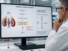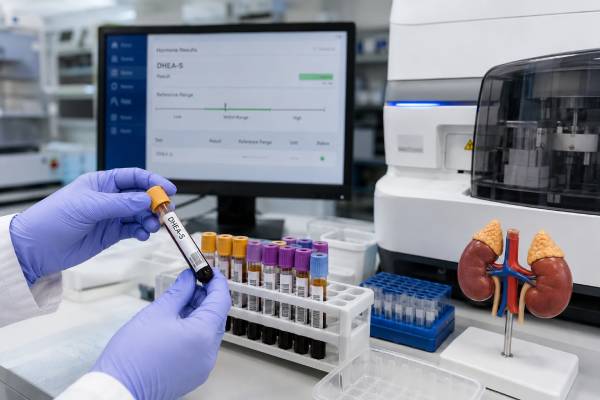
A DHEA-S blood test measures dehydroepiandrosterone sulfate, the most abundant adrenal androgen in circulation. The adrenal glands make DHEA and add a sulfate group to create DHEA-S, a form that remains in the blood longer and varies less from hour to hour. Doctors commonly use it to investigate excess facial or body hair, acne, irregular periods, virilization, unusually early puberty, or a possible adrenal source of androgen excess. DHEA-S also falls when ACTH stimulation or adrenal androgen production is reduced, but a low value alone cannot diagnose adrenal insufficiency. Results depend heavily on age, sex, pubertal stage, laboratory method, medicines, and supplements. Levels are naturally highest in young adulthood and decline over time. Interpretation therefore requires the reference interval on the report, the size of the abnormality, the speed and pattern of symptoms, and related tests such as testosterone, androstenedione, 17-hydroxyprogesterone, cortisol, and ACTH.
- DHEA-S is a stable marker of adrenal androgen production because it has a long half-life and little daily fluctuation.
- High DHEA-S may occur with PCOS, congenital adrenal hyperplasia, supplements, or an androgen-secreting adrenal tumor.
- Low DHEA-S is common with aging and can also occur with glucocorticoid use, pituitary disease, or adrenal insufficiency.
- The normal range changes sharply by age and sex, so a single universal cutoff is not appropriate.
- Rapid virilization or a markedly elevated result needs prompt evaluation, even though most mild elevations are not caused by cancer.
Table of Contents
- DHEA-S and Adrenal Androgen Production
- When the DHEA-S Test Is Useful
- How to Prepare for Testing
- Normal DHEA-S Ranges by Age and Sex
- What High DHEA-S Can Mean
- What Low DHEA-S Can Mean
- Interpreting DHEA-S With Other Tests
- Repeat Testing, Imaging, and Treatment
DHEA-S and Adrenal Androgen Production
DHEA-S is a sulfated steroid made mainly in the zona reticularis, the innermost layer of the adrenal cortex. Pituitary ACTH helps stimulate its production. The molecule itself has relatively weak androgen activity, but tissues can remove the sulfate group and convert DHEA into androstenedione, testosterone, dihydrotestosterone, or estrogens. This local conversion is one reason that symptoms do not always match the blood concentration exactly.
More than 90% of circulating DHEA-S comes from the adrenal glands. That makes it more useful than testosterone for identifying a likely adrenal contribution to androgen excess. Testosterone is produced directly by the ovaries or testes and can also be formed from adrenal precursors, while androstenedione comes from both adrenal and gonadal tissue.
DHEA-S differs from unsulfated DHEA in several practical ways. It circulates at far higher concentrations, remains in the blood for many hours, and shows little of the sharp morning-to-evening change seen with DHEA. A random daytime DHEA-S sample is therefore usually easier to interpret. The DHEA blood test can add detail in specialized steroid evaluations, but DHEA-S is generally the preferred first marker of adrenal androgen output.
The test cannot reveal how much hormone is active inside a specific tissue. It also cannot, by itself, diagnose polycystic ovary syndrome, congenital adrenal hyperplasia, an adrenal tumor, or adrenal insufficiency. Its role is to show whether adrenal androgen production appears appropriate for the person’s age and sex and whether the broader pattern warrants follow-up.
DHEA-S should not be treated as a direct score of vitality, sexual function, immune function, or “adrenal health.” Concentrations decline normally after early adulthood. An older adult may have a much lower level than a 25-year-old while still having normal adrenal cortisol function. Laboratories that advertise a narrow “optimal” range may use criteria that differ from clinical reference intervals and established diagnostic pathways.
Most routine laboratories use an automated immunoassay, while specialty laboratories may use LC-MS/MS. Assay differences can be important near a cutoff or at very low concentrations. When a result is surprising or will lead to imaging, repeat measurement with a reliable method may be appropriate.
When the DHEA-S Test Is Useful
DHEA-S is commonly ordered when there are signs of androgen excess. In women and girls, these may include coarse facial or body hair, persistent acne, scalp hair thinning, irregular or absent menstrual periods, infertility, or virilization. Virilization describes stronger androgen effects such as voice deepening, increased muscle mass, clitoral enlargement, or rapidly progressing hair growth. The faster and more severe the changes, the more urgently clinicians look for a strong androgen source.
The test often helps distinguish an adrenal contribution from an ovarian one. A high DHEA-S points toward adrenal androgen production, although common disorders such as PCOS can include both ovarian and adrenal features. A normal DHEA-S does not rule out PCOS, because many affected people have normal adrenal androgen levels and elevated testosterone or clinical symptoms instead.
In children, DHEA-S may be measured for early pubic or underarm hair, body odor, acne, rapid linear growth, or advanced bone age. DHEA-S begins to rise during adrenarche, often before obvious puberty. Mild isolated premature adrenarche can be benign, but marked values, rapid progression, genital changes, or very young age require evaluation for congenital adrenal hyperplasia, tumors, or exposure to androgen products.
DHEA-S can also contribute to the assessment of:
- An adrenal mass, particularly when imaging features or symptoms raise concern for adrenocortical carcinoma.
- Suspected classic or nonclassic congenital adrenal hyperplasia.
- Unexplained high testosterone or androstenedione.
- Possible adrenal or pituitary insufficiency, as a supportive rather than diagnostic marker.
- Treatment response in selected adrenal androgen disorders.
Testing is less useful as broad screening for fatigue, reduced libido, mood changes, or aging. Those symptoms are common and nonspecific. A low DHEA-S result cannot confirm “adrenal fatigue,” and a normal result cannot exclude sleep disorders, thyroid disease, anemia, depression, medication effects, menopause, or other causes.
A clinician should know how quickly symptoms appeared. Slowly progressive hirsutism beginning around puberty often has a different risk profile from sudden virilization in adulthood. Likewise, a slightly high DHEA-S in a person taking a supplement is interpreted differently from a value several times the upper limit with an enlarging adrenal mass.
How to Prepare for Testing
DHEA-S is measured in serum from a routine venous blood draw. Because the level is comparatively stable through the day, strict morning collection is usually unnecessary. Some clinicians still prefer morning testing when DHEA-S is being drawn with cortisol, ACTH, testosterone, or other hormones that have time-dependent ranges.
Fasting is not always required. Follow the laboratory instructions, especially when the same order includes glucose, insulin, lipids, or another fasting test. Try to use similar conditions for repeat measurements so that changes are less likely to reflect timing or temporary illness.
Tell the clinician and laboratory about all prescription medicines, over-the-counter products, and supplements. DHEA-containing products are especially important. They may be marketed for menopause, fertility, bodybuilding, anti-aging, sexual function, or “adrenal support.” Some combination products list DHEA only in small print. Exposure to another person’s topical testosterone can also matter if skin contact occurs before the product dries.
Glucocorticoids can suppress ACTH and lower DHEA-S. Relevant products include oral prednisone or dexamethasone, injected steroids, high-dose inhaled therapy, potent topical steroids, and repeated joint injections. Oral contraceptives, estrogen therapy, testosterone, antiandrogens, antiseizure medicines, and fertility treatments may also change the clinical picture or related hormone results.
Do not stop a prescribed glucocorticoid without medical instructions. Abrupt withdrawal after sustained treatment can cause adrenal crisis. When drug interference is possible, the clinician may interpret the result in context, adjust timing, or plan a supervised medication strategy rather than simply withholding treatment.
Biotin can interfere with some hormone immunoassays. The effect depends on the platform and dose. A laboratory may recommend stopping high-dose biotin for a defined period before collection, but this should be confirmed rather than guessed.
Menstrual-cycle timing is not usually essential for DHEA-S because production is predominantly adrenal. However, a combined androgen evaluation may be scheduled in the early follicular phase to standardize ovarian hormone measurements. Pregnancy and postpartum physiology require separate interpretation.
Acute illness, major sleep loss, intense exercise, and severe stress can alter adrenal signaling and other test results. A mild unexpected abnormality during temporary illness may be repeated after recovery. The blood draw itself carries only the usual small risks of bruising, soreness, or faintness.
Normal DHEA-S Ranges by Age and Sex
DHEA-S has one of the strongest age patterns among routine hormone tests. Levels are low in early childhood, rise with adrenarche, peak in the second or third decade of life, and then decline gradually. Sex differences are present, and pediatric laboratories may divide ranges by Tanner stage rather than age alone.
Reference intervals vary by assay and population. One laboratory may report micrograms per deciliter, while another uses micromoles per liter. The number cannot be interpreted without its unit. A result of 200 mcg/dL is not equivalent to 200 micromol/L, and DHEA-S values should not be compared with DHEA values.
Illustrative adult intervals from major laboratories often show the following broad pattern:
| Life stage | Expected pattern | Interpretive caution |
|---|---|---|
| Childhood before adrenarche | Low | Use age- and pubertal-stage ranges |
| Adolescence | Rising | Pubertal timing differs widely |
| Young adulthood | Highest average values | Upper limits differ by sex and assay |
| Middle adulthood | Gradual decline | Compare with the current age band |
| Older adulthood | Often substantially lower | Low levels may be physiologic |
For example, some assays list adult female ranges in the low hundreds of mcg/dL during the twenties and much lower upper limits after age 60. Male ranges are often higher at younger ages, then also fall. These are patterns, not replacement values for the report. Even within one age band, reference intervals can be broad.
A mildly out-of-range result is not automatically disease. Reference intervals usually describe the central distribution in a selected population; healthy people may fall just outside. Biological variation and analytical variation also contribute. The farther a value is from the limit, the more consistent it is on repeat testing, and the more closely it matches symptoms, the more weight it carries.
“Optimal” ranges found online are not standardized medical cutoffs. Raising an older adult’s DHEA-S to a young-adult level has not been established as a general health goal. Treatment decisions should be based on a diagnosed condition and meaningful outcomes rather than a target chosen from a wellness chart.
For tumor evaluation, clinicians sometimes use a level around 600–700 mcg/dL as a reason for heightened concern in an adult woman, particularly with rapid virilization. This is not a universal diagnostic threshold. Assay limits, age, menopausal status, symptom speed, testosterone, imaging, and local guidelines all influence the next step. A tumor can occasionally produce a lower value, and a non-tumor condition can produce a high one.
What High DHEA-S Can Mean
A high DHEA-S result indicates increased adrenal androgen exposure or an external source of DHEA. The most straightforward explanation is supplement use. DHEA tablets, sublingual products, creams, and compounded hormones can all raise the level. The exact rise varies by dose, product quality, absorption, and timing.
PCOS is a common cause of mild to moderate androgen elevation in reproductive-age women. DHEA-S may be high, normal, or occasionally low. Diagnosis is not based on this test alone; clinicians consider ovulatory dysfunction, clinical or biochemical androgen excess, and ovarian morphology while excluding thyroid disease, high prolactin, nonclassic congenital adrenal hyperplasia, and other mimics.
Congenital adrenal hyperplasia can increase adrenal androgen production because an enzyme block redirects steroid precursors. The common 21-hydroxylase form is screened with 17-hydroxyprogesterone. Less common enzyme defects produce different steroid patterns. The 17-hydroxyprogesterone test and an androstenedione blood test are often more informative than simply repeating DHEA-S many times.
An androgen-secreting adrenal adenoma or adrenocortical carcinoma is uncommon. Concern rises when DHEA-S is markedly elevated, symptoms progress quickly, several adrenal steroids are abnormal, or imaging shows a large or suspicious lesion. Adrenocortical carcinoma may secrete a mixture of DHEA-S, testosterone precursors, cortisol, or other steroids. A normal cortisol result does not exclude an androgen-secreting lesion.
In children, high DHEA-S can accompany premature adrenarche, early puberty, congenital adrenal hyperplasia, or rarely a tumor. Clinicians compare the result with chronological age, bone age, growth velocity, genital development, and other hormones. Mild pubic hair without rapid growth may require monitoring, while rapid virilization needs prompt specialist assessment.
A high result can also reflect assay interference or a mismatched reference range. Before ordering imaging, clinicians often verify the unit, age band, supplement history, and laboratory method. Repeating a surprising result with LC-MS/MS can be useful when the first measurement was made by immunoassay.
Signs that deserve timely medical review include:
- Voice deepening, clitoral enlargement, or rapid muscle changes.
- Rapidly worsening facial or body hair over a few months.
- Severe new acne with menstrual disruption.
- A very high value confirmed on repeat testing.
- An adrenal mass plus androgen abnormalities.
- Rapid growth, advanced bone age, or genital changes in a child.
The result’s magnitude guides urgency, but it never replaces the full assessment. A scan should answer a well-supported biochemical question, not compensate for incomplete history or unconfirmed testing.
What Low DHEA-S Can Mean
Low DHEA-S is often an expected feature of aging. Levels may fall to a fraction of young-adult values without indicating adrenal failure. Menopause, chronic illness, frailty, and some medicines can further lower the concentration. Because the symptom overlap is broad, low DHEA-S should not automatically be blamed for fatigue, low libido, poor concentration, or reduced well-being.
Glucocorticoid treatment commonly lowers DHEA-S by reducing pituitary ACTH. The effect can occur with oral, injected, inhaled, or potent topical steroids, depending on total exposure. A low result may therefore provide evidence of ACTH suppression but cannot show whether cortisol production is adequate during illness or after the medication is reduced.
Primary adrenal insufficiency damages the adrenal cortex and can lower DHEA-S along with cortisol and aldosterone. Secondary or tertiary adrenal insufficiency reduces ACTH stimulation and can also lower DHEA-S. A low age-adjusted DHEA-S may support suspicion, and a normal value can sometimes make adrenal insufficiency less likely in selected patients, but neither result is definitive.
True adrenal insufficiency is assessed with clinical features and more direct tests. Symptoms may include persistent weakness, weight loss, nausea, abdominal discomfort, low blood pressure, dizziness, salt craving, or recurrent low sodium. Primary disease can also cause high potassium and skin darkening. A cortisol, ACTH, renin, and aldosterone panel helps define the pattern, with stimulation testing when baseline results are uncertain.
Pituitary disorders can reduce ACTH and other hormones. A low DHEA-S may appear alongside low thyroid hormone, low testosterone or estrogen, menstrual changes, infertility, headaches, or visual-field symptoms. Relevant history includes pituitary surgery, radiation, head injury, tumors, and severe postpartum bleeding.
Low DHEA-S is not a reason to self-treat. DHEA supplementation may change testosterone and estrogen exposure and can cause acne, hair changes, mood effects, or problems in people with hormone-sensitive conditions. A clinician should first decide whether there is a disease requiring treatment and whether DHEA would improve an outcome that matters.
A useful interpretation distinguishes three situations: an expected low value for age without concerning symptoms; a medication-related reduction; and a low result that appears with abnormal cortisol, ACTH, electrolytes, or pituitary hormones. Only the third pattern strongly redirects the diagnostic workup.
Interpreting DHEA-S With Other Tests
DHEA-S is rarely the only androgen test needed. The surrounding pattern can help identify the likely source and severity of hormone excess.
| Pattern | Possible interpretation | Typical follow-up |
|---|---|---|
| High DHEA-S, normal or mildly high testosterone | Predominantly adrenal androgen pattern | Review supplements; check 17-hydroxyprogesterone and symptoms |
| Normal DHEA-S, high testosterone | Gonadal source may be more likely | Confirm testosterone and assess ovarian or testicular causes |
| High DHEA-S and high 17-hydroxyprogesterone | Possible congenital adrenal hyperplasia | Specialist testing, often including ACTH stimulation |
| Markedly high multiple adrenal steroids | Strong adrenal steroid production; tumor must be considered | Prompt endocrine review and appropriate imaging |
| Low DHEA-S with low cortisol | Possible adrenal or pituitary insufficiency | ACTH, electrolytes, and dynamic cortisol testing |
Total testosterone should be measured with a method suitable for the low concentrations found in many women and children. Free testosterone is often calculated using total testosterone, sex hormone-binding globulin, and albumin rather than measured with a less reliable direct assay. Androstenedione can reveal additional adrenal or ovarian steroid production.
For suspected nonclassic congenital adrenal hyperplasia, an early-morning 17-hydroxyprogesterone is usually the screening test. Results in an intermediate zone may lead to ACTH stimulation. The exact cutoffs depend on assay and unit.
Cortisol testing is added when there are features of cortisol excess or an adrenal mass. An adrenal lesion can secrete more than one hormone. Conversely, low DHEA-S in a person with possible adrenal failure should be paired with morning cortisol and ACTH rather than interpreted in isolation.
Imaging cannot determine hormone secretion by appearance alone. A benign-looking adrenal adenoma may be functional, while an incidental lesion may be unrelated to the androgen result. Biochemical confirmation and clinical context should guide whether CT or MRI is necessary.
Repeat Testing, Imaging, and Treatment
A mildly abnormal DHEA-S result is often repeated after reviewing supplements, medicines, temporary illness, and the reference range. Repeat testing is especially useful when the person has no matching symptoms or when the value is close to the upper or lower boundary. Using the same laboratory helps assess a trend; using a higher-specificity method may be preferable when interference is suspected.
Marked elevation, rapid virilization, or an abnormal result in a young child generally warrants faster referral to endocrinology. Follow-up may include total and free testosterone, androstenedione, 17-hydroxyprogesterone, cortisol testing, and an adrenal steroid profile. Imaging is selected after the biochemical pattern indicates an adrenal source or when a known adrenal mass must be characterized.
Treatment is directed at the diagnosis rather than the DHEA-S number. PCOS management may address menstrual protection, fertility goals, acne, hirsutism, blood pressure, glucose, and lipids. Congenital adrenal hyperplasia treatment depends on the specific enzyme defect and the need to control androgen excess while avoiding excessive glucocorticoid exposure. A hormone-secreting tumor may require surgery and, for carcinoma, multidisciplinary oncology care.
Low DHEA-S due to adrenal insufficiency is not corrected by DHEA alone. Standard treatment replaces deficient glucocorticoids and, in primary disease, mineralocorticoids when needed. Some guidelines allow a monitored DHEA trial in selected women with confirmed adrenal insufficiency and persistent impaired well-being despite optimized conventional therapy. It is not routine, and it should be stopped if no clear benefit appears.
Over-the-counter DHEA can cause acne, oily skin, unwanted hair growth, scalp hair loss, mood or sleep changes, and changes in sex hormone levels. It may be unsafe in pregnancy, breastfeeding, hormone-sensitive cancers, or some liver and psychiatric conditions. Product dose and purity can vary.
Seek urgent care for severe weakness, repeated vomiting, confusion, fainting, very low blood pressure, or severe abdominal pain in someone at risk for adrenal insufficiency. Those symptoms may indicate adrenal crisis and should never wait for a DHEA-S repeat test.
References
- Society for Endocrinology Clinical Practice Guideline for the Evaluation of Androgen Excess in Women 2025 (Guideline)
- Dehydroepiandrosterone Sulfate in Diagnosing Mild Autonomous Cortisol Secretion and Adrenal Insufficiency 2025
- Evaluating the diagnostic accuracy of androgen measurement in polycystic ovary syndrome: a systematic review and diagnostic meta-analysis to inform evidence-based guidelines 2025 (Systematic Review)
- Age- and sex-adjusted reference intervals for steroid hormones measured by LC-MS/MS using a widely available kit 2023
- DHEA Sulfate Test 2024
- Dehydroepiandrosterone Sulfate, Serum 2026
Disclaimer
This article provides general education and cannot identify the cause of an individual DHEA-S result. Normal ranges differ by age, sex, pubertal stage, units, and laboratory method, so review the report with a qualified clinician. Do not start DHEA or stop steroid treatment without medical guidance.