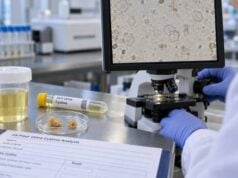
A recurrent kidney stone test panel looks for the reasons stones keep forming and identifies risk factors that can be changed. It is not one standardized laboratory package. A complete evaluation usually combines stone analysis, blood tests, urinalysis, and one or two 24-hour urine collections, then matches the findings to the person’s stone type and medical history. The goal is to move from general advice to targeted prevention. A patient with high urine calcium needs a different plan from someone whose main problem is low urine volume, low citrate, acidic urine, cystinuria, or an infection stone. Recurrent stone formers also deserve testing for systemic conditions that may be missed if only the stone is treated, including primary hyperparathyroidism, renal tubular acidosis, gout-related uric acid disease, bowel-related hyperoxaluria, and chronic urinary infection. The best panel therefore answers two questions: what is the stone made of, and what blood or urine abnormality is driving it?
- A recurrent stone work-up usually includes stone analysis, blood chemistry, urinalysis, and 24-hour urine testing rather than a single “kidney stone blood test.”
- Two 24-hour urine collections are often preferred for high-risk or recurrent stone formers because calcium, sodium, citrate, oxalate, and volume can vary from day to day.
- Key blood tests commonly include calcium, creatinine/eGFR, electrolytes, bicarbonate, uric acid, phosphorus, and PTH when a parathyroid cause is possible.
- Stone analysis is important even when a prior stone type is known: composition can change and can reveal cystine, uric acid, infection, or calcium phosphate disease.
- The panel is most useful on a usual diet after the acute stone episode has settled: severe illness, unusual fluid intake, fasting, or infection can distort the metabolic picture.
Table of Contents
- What a recurrent kidney stone test panel includes
- Blood tests and what they look for
- 24-hour urine testing for recurrent stones
- Stone analysis, urinalysis, and culture
- How the results fit together
- Timing, repeat testing, and collection quality
- What happens after the panel
What a recurrent kidney stone test panel includes
A recurrent stone evaluation is designed to identify both the crystal type and the metabolic environment that allowed it to form. The most useful panel is therefore layered rather than limited to one specimen.
A typical work-up includes:
- a detailed history of prior stone episodes, procedures, imaging, family history, medications, diet, fluid intake, bowel disease, urinary infections, and relevant medical conditions;
- laboratory analysis of a passed or surgically removed stone when available;
- blood tests for kidney function, calcium and other minerals, acid-base status, and uric acid;
- urinalysis, including urine pH and infection assessment when appropriate;
- one or two 24-hour urine collections measuring stone promoters and inhibitors;
- imaging to define current stone burden, obstruction, nephrocalcinosis, or silent stones when clinically indicated.
This approach matters because recurrence can have more than one cause. A person with calcium oxalate stones may have hypercalciuria, hyperoxaluria, hypocitraturia, low volume, high sodium intake, or several of these at once. Another patient may repeatedly form uric acid stones primarily because urine pH is persistently low even though the daily uric acid amount is not markedly elevated.
Guidelines generally classify recurrent stone formers as higher risk and support a more detailed metabolic evaluation. Uric acid, cystine, and infection stones are also considered high-risk because they often recur unless the underlying chemistry or infection is corrected.
The work-up should still be individualized. A young patient with a strong family history and multiple bilateral stones may need evaluation for genetic causes. A patient with chronic diarrhea or bariatric surgery may need special attention to oxalate and citrate. A patient with recurrent urinary infection and branching stones needs an infection-focused evaluation rather than only a dietary review.
For a narrower panel focused on urine chemistry, see the kidney stone test panel. Recurrent disease usually adds greater emphasis on stone analysis, systemic causes, and long-term follow-up.
Blood tests and what they look for
Blood tests help find systemic disorders that a 24-hour urine collection cannot diagnose by itself. They also establish whether a proposed prevention medicine is safe.
Calcium and parathyroid hormone
Serum calcium is one of the most important screening tests in recurrent calcium stone disease. Repeated hypercalcemia raises concern for primary hyperparathyroidism, particularly when PTH is elevated or fails to suppress. The PTH test for kidney stones is usually interpreted with calcium, not by itself.
A normal blood calcium does not exclude high urine calcium. Idiopathic hypercalciuria is common and can occur with completely normal serum calcium.
Creatinine and eGFR
Creatinine and estimated glomerular filtration rate assess kidney function. Recurrent stones can coexist with chronic kidney disease, and kidney function affects the safety and dosing of medicines such as potassium citrate, allopurinol, and some diuretics. Reduced filtration also changes how PTH and some urine markers should be interpreted.
Electrolytes and bicarbonate
Sodium, potassium, chloride, and bicarbonate provide clues about fluid balance and acid-base disorders. A low bicarbonate level can suggest metabolic acidosis. In a stone former with alkaline urine and low citrate, that pattern can raise concern for distal renal tubular acidosis, a condition associated with calcium phosphate stones and nephrocalcinosis.
Uric acid
Serum uric acid helps evaluate gout, hyperuricemia, and uric acid metabolism. A high blood level is not required for uric acid stones, because persistently acidic urine is often the more important driver. Still, the blood result adds context and can influence treatment when gout or urate overproduction is present.
Phosphorus and other tests
Phosphorus can support interpretation of parathyroid and mineral disorders. Depending on the history, clinicians may also check magnesium, vitamin D, complete blood count, liver tests, or other targeted studies. No single fixed blood panel is correct for every recurrent stone former.
The key principle is that blood tests screen for causes that would change management. A high serum calcium result may lead to endocrine treatment. A low bicarbonate may redirect the evaluation toward acid-base disease. Reduced kidney function may alter drug choices even if the urine profile otherwise suggests standard therapy.
24-hour urine testing for recurrent stones
The 24-hour urine collection is the core metabolic test for many recurrent stone formers because it measures what the kidneys excrete over a complete day. A comprehensive panel commonly includes urine volume, calcium, oxalate, citrate, uric acid, sodium, potassium, magnesium, phosphorus, creatinine, urea, and pH. Many laboratories also calculate supersaturation for calcium oxalate, calcium phosphate, and uric acid.
Two consecutive collections are often recommended because stone risk factors vary with meals, work schedules, hydration, physical activity, and salt or protein intake. If one day shows 1.4 L of urine and the next shows 2.3 L, or sodium and calcium differ substantially, the variation itself is clinically useful.
The most important measurements include:
- Urine volume: low volume concentrates every stone-forming solute. Prevention plans commonly aim for more than 2.0–2.5 L of urine per day, with higher targets for some disorders.
- Urine calcium: higher excretion raises calcium stone risk, especially when sodium is high or volume and citrate are low.
- Urine oxalate: elevated oxalate can reflect diet, vitamin C exposure, intestinal malabsorption, bariatric surgery, or rare primary hyperoxaluria.
- Urine citrate: citrate binds calcium and inhibits crystal formation. Low citrate is common with metabolic acidosis, chronic diarrhea, high dietary acid load, and low potassium states.
- Urine uric acid: excess excretion can contribute to uric acid and some calcium stone patterns.
- Urine pH: low pH favors uric acid, while higher pH can favor calcium phosphate; very alkaline urine with infection raises concern for struvite.
- Urine sodium: helps estimate salt exposure and is important because higher sodium intake often increases urine calcium.
- Creatinine: helps judge whether the collection was plausibly complete for that individual.
A 24-hour urine stone risk profile is especially useful when the goal is to see these relationships together rather than chasing isolated cutoffs.
An abnormal result should also be judged by whether it is reproducible and biologically plausible. A single day with unusually salty food can raise urine sodium and calcium, while heavy exercise, hot weather, or diarrhea can sharply reduce volume. If one collection shows a striking abnormality that is absent on the second collection, the clinician may review the collection diary, creatinine, diet, and timing before committing to long-term medication. The reverse is also important: a value that looks only mildly abnormal on both days may be more convincing than one extreme value on an atypical day. Repeated patterns help separate persistent physiology from noise.
This is one reason treatment targets are usually tied to the whole profile and to stone activity. A modest fall in calcium or oxalate can be meaningful if supersaturation falls and no new stones form, while a laboratory value that reaches the printed reference range may still be insufficient if stones continue to grow.
Stone analysis, urinalysis, and culture
Stone analysis answers a different question from blood and urine chemistry: it identifies what the stone is actually made of. For recurrent stones, this is high-value information because composition guides which abnormalities deserve the most attention.
Modern stone analysis typically uses infrared spectroscopy or X-ray diffraction. Results may identify calcium oxalate, calcium phosphate, uric acid, struvite, cystine, or mixed components. Chemical analysis alone is less informative than these physical methods.
A kidney stone analysis test should be performed when a stone is available, particularly if no prior composition is known. Repeat analysis can also be useful when the recurrence pattern changes or prevention is not working. A patient who previously formed calcium oxalate stones can later form calcium phosphate or another composition, especially after changes in urine pH or treatment.
Routine urinalysis adds information about blood, white cells, crystals, protein, specific gravity, and urine pH. Microscopy may reveal characteristic crystals. Hexagonal cystine crystals strongly suggest cystinuria, while abundant struvite-type crystals in alkaline urine may support an infection-stone work-up.
Urine culture is especially important when recurrent stones occur with fever, pyuria, recurrent urinary tract infection, alkaline urine, staghorn calculi, or suspected struvite. Infection stones form in association with urease-producing organisms and usually require both control of infection and removal of stone material.
A negative routine urinalysis between episodes does not rule out a metabolic stone disorder. Likewise, finding bacteria in one sample does not prove that the stone is an infection stone. The stone composition, culture history, imaging pattern, and urine chemistry need to agree.
How the results fit together
The value of a recurrent stone panel comes from combining results into a pattern. Several common examples show why this matters.
High urine calcium plus high urine sodium
This suggests that sodium intake may be increasing calcium excretion. The prevention plan may emphasize sodium reduction while maintaining normal dietary calcium. If hypercalciuria persists, a clinician may discuss a thiazide-type medicine depending on the full risk profile and current evidence.
Low citrate plus low bicarbonate or chronic diarrhea
This pattern points toward alkali loss or metabolic acidosis. Potassium citrate may be appropriate in selected patients, but the cause should be identified rather than treating the urine number alone.
High oxalate after bowel surgery
Enteric hyperoxaluria becomes more likely in a patient with fat malabsorption, inflammatory bowel disease, short bowel, or certain bariatric procedures. Management differs from ordinary dietary hyperoxaluria and may include calcium with meals, dietary fat and oxalate changes, and focused gastrointestinal care.
Low urine pH with uric acid stones
Persistently acidic urine is often the dominant abnormality. In that setting, the uric acid stone risk profile may show that raising urine pH is more important than aggressively lowering total uric acid excretion.
High calcium with non-suppressed PTH
This combination suggests a parathyroid cause rather than simple idiopathic hypercalciuria. Treating primary hyperparathyroidism can address a systemic driver of stones, bone loss, and hypercalcemia.
Positive cystine testing
Cystinuria changes the prevention target substantially. Patients often need very high urine volume, urine alkalinization, sodium restriction, and sometimes cystine-binding medication. A general calcium-stone plan is not sufficient.
This pattern-based approach is also why a normal result in one category does not end the evaluation. A person can have normal urine calcium and still form stones because volume is low, oxalate is high, citrate is low, or urine pH is unfavorable.
Timing, repeat testing, and collection quality
A metabolic panel should reflect the person’s usual life as closely as possible. Collecting urine during severe pain, vomiting, fasting, hospitalization, an active infection, or unusually aggressive hydration can produce values that are hard to apply to everyday prevention.
Guidelines generally recommend waiting until the acute episode has resolved and the patient is back in the usual environment, diet, and fluid pattern. Exact timing varies. European guidance suggests that specific metabolic testing is best performed once stable and preferably around three months after an acute episode, while other guidance takes a pragmatic approach and emphasizes not delaying evaluation unnecessarily when the patient has recovered.
During the collection, every void matters. Missing urine usually lowers measured 24-hour excretion and can make calcium, oxalate, citrate, sodium, uric acid, and volume look falsely reassuring. Overcollection or extending the time well beyond 24 hours can have the opposite effect. Creatinine excretion provides a rough collection-quality check, but it does not identify every error.
Do not deliberately change the diet to “pass” the test. A low-sodium, high-fluid day that is very different from normal habits may hide the risk factors the clinician needs to see. The baseline collection is often most useful when it captures ordinary intake.
After a prevention plan is started, repeat testing shows whether the intervention worked. European guidance suggests a first follow-up 24-hour urine about 8–12 weeks after pharmacologic prevention begins, with further adjustment if risk factors remain abnormal. Once values are stable, yearly testing is often reasonable for higher-risk patients, although frequency depends on recurrence, stone burden, medication, and clinical judgment.
A repeat urine supersaturation test can be especially useful because a good intervention should improve the chemical tendency to crystallize, not merely move one isolated marker.
What happens after the panel
The next step is to convert the abnormal pattern into a prevention plan that can be measured. General measures usually include enough fluid to produce high urine volume, normal food-based calcium intake, moderation of sodium, and avoiding excessive animal protein. Those recommendations are then modified according to stone type and urine chemistry.
Examples of targeted treatment include potassium citrate for selected patients with hypocitraturia or uric acid stones, thiazide-type therapy for selected recurrent calcium stone formers with persistent hypercalciuria, allopurinol in specific hyperuricosuric settings, and cystine-binding drugs for cystinuria that remains uncontrolled with fluid, diet, and alkalinization. Infection stones require a different strategy centered on complete stone management and microbiologic control.
A prevention plan should include a measurable goal. Instead of “drink more,” the goal might be urine volume above 2.5 L/day. Instead of “eat less salt,” follow-up urine sodium can show whether sodium exposure actually fell. Instead of assuming citrate therapy worked, repeat urine citrate, pH, potassium, and supersaturation can verify the effect.
Recurrent stone testing also helps avoid incorrect treatment. A patient with calcium stones should not automatically be told to eliminate dietary calcium, because normal calcium intake can reduce intestinal oxalate absorption. A patient with uric acid stones may need urine alkalinization even when serum uric acid is not severely elevated. A patient with hypercalcemia should not be managed as routine idiopathic hypercalciuria until a parathyroid cause is excluded.
Finally, prevention does not replace urgent care. Fever or chills with an obstructing stone, inability to urinate, severe uncontrolled pain, or persistent vomiting can signal a urologic emergency. Metabolic testing is for preventing the next stone; acute obstruction or infection requires immediate clinical management.
A well-designed recurrent kidney stone panel therefore does more than generate a long list of laboratory values. It links stone composition, blood chemistry, urine chemistry, infection status, and collection quality into a specific explanation for recurrence and a plan that can be tested again over time.
References
- Metabolic Evaluation and Recurrence Prevention – EAU Guidelines on Urolithiasis – Uroweb 2026 (Guideline)
- Metabolic evaluation for prevention of kidney stones – CARI Guidelines 2025 (Guideline)
- Metabolic evaluation of urolithiasis: a narrative review. 2025 (Review)
- 24-Hour Urinary Chemistries and Kidney Stone Risk 2024
Disclaimer
This article is for general education and does not replace a personalized stone evaluation. The tests needed, treatment thresholds, and follow-up schedule depend on stone composition, kidney function, medications, and individual risk. Seek urgent medical care for a stone accompanied by fever, inability to urinate, severe uncontrolled pain, or persistent vomiting.