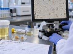
A struvite stone test is not one single laboratory test. Struvite stones are infection stones, so diagnosis usually combines stone analysis, urine culture, urine pH, urinalysis, imaging, and sometimes metabolic testing after the infection and stone have been treated. Struvite is magnesium ammonium phosphate. It forms when urease-producing bacteria split urea, generating ammonia and making the urine strongly alkaline. The most classic organism is Proteus, but other urease-producing bacteria can also be involved. A high urine pH or positive culture alone does not prove a struvite stone; direct stone analysis is the strongest way to identify its composition. These stones matter because they can grow quickly, branch through the kidney as staghorn stones, harbor bacteria, and contribute to recurrent infection or sepsis. Testing therefore aims to identify both the stone and the infection that sustains it, then confirm that the infection and residual stone burden are controlled.
- Struvite is an infection stone made mainly of magnesium ammonium phosphate and forms in alkaline urine created by urease-producing bacteria.
- Stone analysis is the most direct test: urine pH and culture support the diagnosis but cannot identify stone composition by themselves.
- Urine pH is often markedly alkaline: struvite precipitation is favored above about pH 7.2, although pH varies over time and after antibiotics.
- Proteus species are classic causes, but Morganella, Providencia, some Klebsiella, Staphylococcus, and other urease-producing organisms can also be involved.
- Fever, chills, severe flank pain, confusion, or low blood pressure with an obstructing stone can signal infected obstruction or sepsis and needs urgent medical care.
Table of Contents
- What struvite stones are and why infection matters
- Tests used to diagnose a struvite stone
- Urine pH, bacteria, and culture results
- Stone analysis and imaging findings
- How struvite test results are interpreted
- Treatment and follow-up testing
- Common pitfalls and urgent warning signs
What struvite stones are and why infection matters
Struvite stones form because of a specific chemical effect of urinary infection. Urease-producing bacteria release the enzyme urease, which breaks urea into ammonia and related products. The process raises urine pH and increases ammonium. In that alkaline environment, magnesium, ammonium, and phosphate can crystallize as struvite. Carbonate apatite is also common in infection stones.
This mechanism is different from the usual calcium oxalate stone, which can form without infection. A person may have bacteria in the urine and still form a non-struvite stone. Conversely, a stone may contain a mixture of struvite and metabolic components, suggesting that an existing calcium stone later became infected.
Struvite stones are considered high risk because the bacteria and stone can reinforce each other. The stone may act as a reservoir for organisms, while ongoing urease activity keeps the urine favorable for further crystal growth. Some infection stones enlarge into staghorn calculi that occupy a large portion of the renal collecting system.
Risk is higher when the urinary tract does not drain or empty normally. Predisposing factors include neurogenic bladder, spinal cord injury, urinary diversion, indwelling catheters, foreign bodies, urethral obstruction, bladder diverticula, and ureteropelvic junction obstruction. These conditions can promote persistent or recurrent infection.
Struvite disease therefore needs more than a routine “kidney stone diet.” The core problem is infection plus stone material. Metabolic abnormalities can coexist, especially in mixed stones, but the infection must be addressed first.
Tests used to diagnose a struvite stone
A struvite evaluation usually combines several tests because no single urine marker is diagnostic.
Stone analysis
If a stone has been passed or removed, laboratory analysis can identify struvite, carbonate apatite, ammonium urate, calcium oxalate, calcium phosphate, or a mixture. Infrared spectroscopy or X-ray diffraction provides a direct composition result. A kidney stone analysis test is especially important when infection stones are suspected because treatment and recurrence risk differ from ordinary calcium stones.
Urinalysis and urine pH
Urinalysis may show alkaline pH, white blood cells, bacteria, blood, and crystals. A urine pH test for kidney stones can support the pattern, but pH should be repeated because it changes during the day and can fall after antibiotics or other treatment.
Urine culture
Culture identifies bacteria growing in the urine and provides antibiotic susceptibility information. It is central to managing infection, but a midstream culture may not perfectly represent bacteria living inside the kidney or stone.
Imaging
Ultrasound or CT can show stone size, location, branching staghorn configuration, hydronephrosis, and residual fragments. Imaging cannot prove struvite composition, but a large branching stone in a patient with recurrent alkaline urinary infection raises strong suspicion.
Blood tests
Creatinine and eGFR assess kidney function. A complete blood count, inflammatory markers, electrolytes, and blood cultures may be used when systemic infection is suspected. In an acute infected obstruction, these tests help assess severity rather than stone composition.
Once infection is controlled and stone material has been removed, a 24-hour urine evaluation may be appropriate, especially for mixed stones. That can uncover hypercalciuria, low citrate, high oxalate, or another metabolic risk that would otherwise continue to produce non-infection stones.
Urine pH, bacteria, and culture results
Urine pH is one of the strongest clues to an active urease effect. Struvite precipitation is favored in strongly alkaline urine, with European guidance noting that struvite typically precipitates above about pH 7.2. Carbonate apatite can begin to crystallize at a somewhat lower alkaline pH, around 6.8.
That does not mean every urine pH above 7.2 equals a struvite stone. Urine can become alkaline from diet, some medications, delayed sample processing, renal tubular disorders, or contamination. The meaningful pattern is alkaline urine plus infection evidence plus a compatible stone.
The organisms most strongly associated with urease production include:
- Proteus species, particularly Proteus mirabilis;
- Providencia rettgeri and some other Providencia species;
- Morganella morganii;
- Corynebacterium urealyticum;
- Ureaplasma urealyticum;
- some Klebsiella, Serratia, and Staphylococcus species.
Urease production can vary by species and strain. Common organisms such as Escherichia coli are usually not strong urease producers, yet cultures from patients with struvite stones may still grow E. coli or other organisms. This can happen because the current bladder culture does not necessarily identify the organism that initiated stone formation, because mixed infections occur, or because antibiotics altered the microbial picture.
A multicenter study of stone formers found positive midstream cultures in only about two-thirds of patients with struvite stones. That is an important limitation: a negative culture does not completely exclude an infection stone, especially after recent antibiotics.
For a detailed look at how bacterial growth is confirmed, the urine culture test and urine culture and sensitivity testing explain how colony growth and antibiotic susceptibility are interpreted.
Why culture timing and sample quality matter
Culture is most informative when a clean sample is obtained before antibiotics, as long as delaying treatment is medically safe. In a sick patient, antibiotics should not be postponed simply to improve a culture. When treatment has already started, the clinician interprets a negative result with that limitation in mind and may repeat the culture later.
Contamination can also complicate interpretation. A clean-catch midstream specimen reduces skin and genital contamination, but patients with catheters, urinary diversions, or unusual anatomy may require a different sampling method. “Mixed flora” on a culture often means the specimen is not ideal for identifying one causative organism, although the clinical situation determines whether repeat sampling is needed.
The location of the culture matters as well. Bladder urine may differ from urine trapped above an obstruction, and bacteria embedded within a stone may not be represented by a routine midstream sample. For patients undergoing a procedure, urologists may obtain renal pelvis urine or a stone culture. Those results can be particularly useful when prior cultures have been negative or have grown inconsistent organisms.
Because struvite stones are tied to urease activity rather than merely the presence of bacteria, the clinician asks whether the microbiology and chemistry make sense together. A repeated Proteus culture with pH 8.0 and a branching stone is much more persuasive than one isolated alkaline dipstick in an otherwise asymptomatic patient.
A negative midstream culture does not completely exclude an infection stone. Antibiotics given before the sample can suppress bacterial growth, and organisms within a stone or the renal collecting system may not always appear in a bladder specimen. In surgical cases, clinicians may obtain renal pelvic urine or stone culture because those samples can reveal organisms missed by a routine culture. This distinction matters when infection persists despite apparently negative urine testing.
The reverse problem also occurs: a positive culture in a patient with a stone does not prove the stone is struvite. Ordinary calcium stones can coexist with urinary tract infection. Stone composition remains the best way to determine whether infection minerals are actually present, and culture identifies the organism that needs treatment rather than defining the mineral by itself.
Stone analysis and imaging findings
Direct stone analysis carries more diagnostic weight than urine pH alone because it answers the composition question. A result may report pure struvite, struvite plus carbonate apatite, or a mixed stone containing calcium oxalate or calcium phosphate.
Mixed composition can be clinically informative. A stone with a large calcium component plus struvite may have started as a metabolic stone and later become infected. After removal and infection control, the patient may still need a metabolic prevention plan for the calcium component.
Urine microscopy can also provide clues. Struvite crystals are often described as rectangular “coffin-lid” crystals, but crystal appearance is not specific enough to diagnose a stone. Crystals can be present without a formed stone, and a stone can exist even when crystals are not seen in a small urine sample. A separate struvite crystals in urine result is therefore supportive rather than definitive.
Imaging defines the practical burden of disease. CT is highly sensitive for stones and can show branching calculi, obstruction, gas in unusual infection settings, and residual fragments after treatment. Ultrasound avoids radiation and is useful for hydronephrosis and follow-up, but it is less precise for stone composition and small residual fragments.
A staghorn shape is often associated with infection stones, but not every staghorn calculus is struvite. Calcium phosphate, calcium oxalate, uric acid, and mixed stones can also become large and branching. The appearance should therefore trigger analysis, not replace it.
Residual stone fragments are especially important in struvite disease because colonized material can serve as a nidus for regrowth and recurrent infection. The aim of treatment is usually to remove stone material as completely as safely possible.
How struvite test results are interpreted
The most convincing pattern is a stone analysis showing struvite together with evidence of urease-associated urinary infection. Other combinations require more nuance.
| Finding | What it suggests | Important limitation |
|---|---|---|
| Struvite on stone analysis | Confirms an infection-stone component | Does not identify the current organism |
| Urine pH >7.2 with urease-producing bacteria | Strongly supports active chemistry favorable for struvite | Still does not prove an existing stone is struvite |
| Positive culture with normal or mildly acidic pH | UTI may be present without active urease alkalinization | Recent treatment and timing can alter pH |
| Alkaline urine but negative culture | Could reflect prior antibiotics, noninfectious alkalinity, or sampling limits | Repeat culture and stone analysis may be needed |
| Mixed struvite-calcium stone | Infection plus a possible metabolic stone tendency | Metabolic testing is most useful after infection control |
A single dipstick pH should not drive the diagnosis. Freshly measured pH is more reliable because urine can become more alkaline after standing if bacteria proliferate in the sample. Repeated values are more useful than one isolated reading.
The same applies to culture. If a patient recently received antibiotics, a bladder culture may be negative even though the stone was formed in an infected environment. During surgery, renal pelvis or stone cultures can sometimes provide information that differs from the preoperative midstream sample.
If infection has resolved but stones keep recurring, the clinician should look for an additional metabolic cause rather than assuming every new stone is struvite.
Treatment and follow-up testing
The central treatment principle is removal of stone material plus control of infection. Antibiotics alone may suppress bacteria but often cannot sterilize a large colonized stone permanently. Likewise, stone removal without treating persistent bacteriuria leaves the patient vulnerable to recurrence.
For large or complex stones, percutaneous nephrolithotomy is commonly used, sometimes with staged or combined procedures. The exact surgical plan depends on stone size, anatomy, kidney function, infection status, and patient factors.
Antibiotic therapy should be guided by culture and susceptibility when possible. The ideal duration after stone removal is individualized because evidence does not support one universal course for every patient. Persistent bacteriuria needs active treatment and reassessment.
In selected difficult cases with residual stone or persistent urease-producing infection, a urease inhibitor such as acetohydroxamic acid may be considered where available. It can reduce urease activity but has significant adverse effects and is not routine first-line therapy. Urinary acidification strategies are also used in some settings, but they require clinician supervision. The goal is not to make urine as acidic as possible. Acidifying agents can interact with kidney function, electrolyte balance, diet, and other medicines, and the evidence for long-term use is less robust than the evidence supporting stone removal and infection control. Patients should not try to force urine pH down with supplements or large dietary changes without a treatment plan.
Antibiotic selection also deserves caution. The organism, susceptibility pattern, kidney function, allergy history, pregnancy status, and severity of infection all affect the choice and dose. Repeated empiric antibiotic courses without culture can temporarily suppress symptoms while leaving a colonized stone or resistant organism in place. In recurrent cases, the clinical question is not only “Which antibiotic works?” but also “Is there residual stone or obstruction that prevents the infection from clearing?”
Follow-up commonly includes:
- repeat urine culture when infection persistence or recurrence is a concern;
- repeat urinalysis and pH when clinically useful;
- imaging to look for residual or recurrent stones;
- kidney function testing;
- metabolic 24-hour urine testing after stone removal and infection control when the stone is mixed or recurrent.
A recurrent kidney stone test panel can help identify noninfectious factors after the immediate struvite problem is controlled. This prevents a mixed stone former from receiving infection treatment while an untreated calcium or uric acid risk remains.
Common pitfalls and urgent warning signs
One common mistake is assuming that any alkaline urine means struvite. Another is assuming that any UTI in a stone former means the stone was caused by infection. Struvite requires the specific urease-driven chemical environment, and stone composition provides the clearest confirmation.
A second mistake is relying on one negative urine culture. Antibiotics, intermittent bacterial shedding, sampling location, and colonized stone material can all make the bladder culture less informative than expected.
A third mistake is treating only the urine test while leaving a substantial infected stone in place. Infection stones can remain a bacterial reservoir, so complete or near-complete stone clearance is a major part of recurrence prevention.
The most important safety issue is infected obstruction. A stone blocking urine flow while infection is present can rapidly lead to pyelonephritis, sepsis, kidney injury, and shock. Seek urgent medical care for fever or shaking chills with flank pain, confusion, rapid breathing, low blood pressure, marked weakness, inability to pass urine, or persistent vomiting. An infected obstructed kidney may require emergency drainage in addition to antibiotics.
People with indwelling catheters, urinary diversions, neurogenic bladder, recurrent Proteus infections, or known staghorn stones may need closer surveillance even when symptoms are mild. Struvite stones can become large before causing classic renal colic.
The practical goal of testing is therefore broader than labeling a stone “struvite.” It is to document infection-stone composition when possible, identify the organism, assess how alkaline the urine is, define the anatomic stone burden, and verify after treatment that neither infected stone material nor recurrent bacteriuria is continuing the cycle.
References
- Metabolic Evaluation and Recurrence Prevention – EAU Guidelines on Urolithiasis – Uroweb 2026 (Guideline)
- Management of urinary stones by experts in stone disease (ESD 2025) 2025
- Spectrum of Bacterial Pathogens from Urinary Infections Associated with Struvite and Metabolic Stones. 2023
- Metabolic evaluation for prevention of kidney stones – CARI Guidelines 2025 (Guideline)
- Pharmacologic treatment of kidney stones: Current medication and pH monitoring 2023 (Review)
Disclaimer
This article is for general education and does not diagnose an infection stone or provide an antibiotic plan. Struvite evaluation and treatment depend on stone composition, culture results, imaging, kidney function, and whether obstruction is present. Fever or chills with a suspected obstructing stone, confusion, low blood pressure, inability to urinate, or severe illness requires urgent medical care.