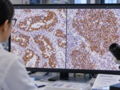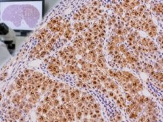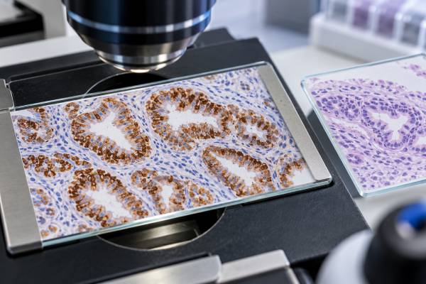
Villin immunohistochemistry (IHC) is a tissue stain used to identify intestinal and gastrointestinal-type differentiation in tumors. Villin is an actin-binding protein concentrated in the brush border of normal intestinal epithelial cells. Many colorectal, gastric, pancreatobiliary, and other gastrointestinal adenocarcinomas express villin, often with apical or brush-border staining. That makes it useful when a pathologist is evaluating the origin of a metastatic adenocarcinoma or a mucinous tumor. Villin is sensitive in many gastrointestinal cancers but is not specific enough to identify a primary site on its own. Large tumor studies have shown expression in renal tumors, mucinous ovarian cancers, yolk sac tumors, neuroendocrine neoplasms, and subsets of other cancers. Some poorly differentiated colorectal cancers can also lose villin. The most reliable interpretation combines villin with morphology and markers such as CDX2, SATB2, CK7, and CK20, plus organ-specific stains chosen for the differential diagnosis.
- Villin positivity usually appears in the cytoplasm and may concentrate at the apical or brush-border surface.
- Many colorectal and other GI adenocarcinomas are villin positive, but positivity is not specific to the gastrointestinal tract.
- SATB2 and CDX2 usually provide more specific lower-GI information than villin alone.
- A negative villin stain does not exclude colorectal cancer, especially in poorly differentiated tumors.
- Villin IHC is interpreted as part of a panel and has no blood reference range or patient preparation.
Table of Contents
- What the villin IHC test detects
- Villin in colorectal and other GI cancers
- What positive villin staining means
- Non-GI tumors that can be villin positive
- Why a GI cancer can be villin negative
- How villin is used in IHC panels
- What the result means for patients
What the villin IHC test detects
Villin is a cytoskeletal protein associated with microvilli and the intestinal brush border. IHC detects the protein in formalin-fixed tissue. In adenocarcinomas, staining may be cytoplasmic, membranous, or concentrated along the luminal/apical surface of glands.
The apical brush-border pattern is particularly suggestive of intestinal differentiation, but the exact pattern varies by tumor and antibody method. Pathologists assess which tumor cells are positive, the distribution, and whether staining fits the microscopic architecture.
Villin IHC is not a serum marker and does not provide a numeric reference interval. Its value is comparative and morphologic: it helps answer whether a tumor is showing a gastrointestinal-type differentiation program and how that finding fits with other tissue markers.
Villin in colorectal and other GI cancers
Colorectal adenocarcinomas frequently express villin, as do many gastric, pancreatic, and biliary adenocarcinomas. Because of this broad distribution, villin is better viewed as a gastrointestinal/intestinal differentiation marker than as a colorectal-specific marker.
When colorectal origin is the main question, CDX2 and SATB2 often provide stronger site-specific information. SATB2 is particularly useful for lower-GI origin, while CDX2 is highly sensitive for intestinal differentiation but can also label upper-GI and selected non-GI tumors.
Villin can still add value when those markers are equivocal or when a broader gastrointestinal phenotype is being tested. A coherent apical villin pattern alongside CDX2 and CK20 may strengthen the interpretation of a metastatic gland-forming carcinoma.
What positive villin staining means
A positive villin result means detectable protein is present in the tumor. Pathologists may describe diffuse or focal cytoplasmic staining and note apical or brush-border accentuation. Strong apical staining in a gland-forming carcinoma can be a useful clue toward intestinal differentiation.
The stain should not be converted into a rigid yes/no primary-site rule. In a mucinous ovarian tumor, for example, villin positivity can occur in both primary ovarian mucinous carcinoma and metastatic gastrointestinal carcinoma. Recent data suggest that pairing villin with SATB2 can improve discrimination because SATB2 is much more specific for lower-GI origin in that setting.
A CK20 IHC stain may add another intestinal-associated signal, while CK7 and PAX8 can support competing ovarian or pancreatobiliary interpretations depending on the case.
Non-GI tumors that can be villin positive
Villin expression is not confined to gastrointestinal cancers. Large tissue microarray studies have documented positivity in renal tumors, mucinous ovarian cancers, yolk sac tumors, neuroendocrine neoplasms, and additional carcinomas. Some primary lung adenocarcinomas, particularly mucinous tumors or tumors with prominent microvilli, can also express villin.
This is a major pitfall when a villin-positive tumor is found in the lung. The differential may include a primary pulmonary mucinous adenocarcinoma and metastatic colorectal carcinoma. CK7, SATB2, CDX2, TTF-1, Napsin A, mucin markers, imaging, and clinical history are more informative together than villin alone.
Villin positivity can therefore support a differentiation phenotype without proving anatomy. The more unusual the location, the more important it is to demand agreement from several independent lines of evidence.
Why a GI cancer can be villin negative
Not every colorectal or gastrointestinal carcinoma retains villin. Poorly differentiated colorectal cancers and tumors with microsatellite instability may show reduced or absent expression in some series. Advanced tumors can also lose mature brush-border differentiation.
Technical issues can mimic biologic loss. Necrosis, fixation problems, very small samples, and heterogeneity may reduce staining. If the morphology and clinical findings strongly favor colorectal origin, a negative villin result should prompt correlation with SATB2, CDX2, CK20, and other markers rather than immediate rejection of the diagnosis.
This limitation illustrates why pathologists prefer panels. A highly differentiated tumor may express several intestinal markers strongly, while a high-grade metastasis may retain only one or two.
How villin is used in IHC panels
For a suspected lower-GI metastasis, a practical panel may include SATB2, CDX2, CK20, and CK7, with villin as an additional intestinal differentiation marker. For pancreatobiliary tumors, CK7, MUC markers, SMAD4, and clinical imaging may be more central. For ovarian mucinous tumors, PAX8 and the CK7/SATB2 balance can be particularly helpful.
Villin also helps in selected differentials involving bladder adenocarcinoma, cholangiocarcinoma, lung adenocarcinoma, and metastatic gastrointestinal cancer. The marker’s broad sensitivity becomes useful when the question is “Does this tumor show intestinal differentiation?” rather than “Exactly which organ did it come from?”
The panel is always shaped by morphology. A MUC2 IHC stain may add evidence of intestinal-type mucin production in a mucinous tumor but, like villin, must be interpreted in context.
What the result means for patients
Villin IHC is primarily a diagnostic aid. It usually does not determine treatment directly and is not used to monitor cancer in blood. Its value is helping the pathology team classify a tumor and identify the most likely primary site.
If a report says “villin positive,” look for the other stains and the pathologist’s integrated comment. Ask whether the pattern was apical or diffuse, whether SATB2 and CDX2 supported lower-GI origin, whether CK7 suggested another site, and whether imaging agrees with the proposed primary.
Once tumor origin is established, stage and molecular biomarkers—not villin intensity—usually drive treatment decisions. In colorectal cancer, for example, mismatch-repair status, RAS/BRAF alterations, HER2 in selected cases, and other molecular features can be more clinically actionable.
References
- Evaluation of Diagnostic Accuracy of Immunohistochemical Markers of SATB2 and Villin in Differential Diagnosis of Ovarian and Gastrointestinal Mucinous Carcinoma 2025 (Study)
- The utility of the lineage specific immunohistochemical stains SATB2, CDX2, and villin, and the mucin glycoproteins MUC2, MUC5AC, and MUC6 to distinguish pulmonary invasive mucinous adenocarcinoma from metastatic colorectal carcinoma 2024 (Study)
- Villin expression in human tumors: a tissue microarray study on 14,398 tumors. 2022 (Study)
- Loss of villin immunoexpression in colorectal carcinoma is associated with poor differentiation and survival 2013 (Study)
- Distribution and pattern of expression of villin, a gastrointestinal-associated cytoskeletal protein, in human carcinomas: a study employing paraffin-embedded tissue 1991 (Study)
Disclaimer
Villin IHC is an ancillary tissue stain and cannot establish gastrointestinal or colorectal origin by itself. Results must be interpreted with morphology, companion markers, clinical history, and imaging. Some non-GI tumors are positive and some GI cancers are negative.