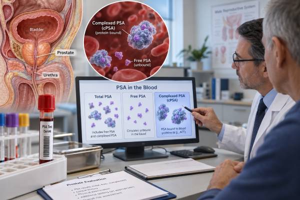
Complexed prostate-specific antigen (complexed PSA or cPSA) measures the portion of PSA in blood that is bound to certain proteins, especially alpha-1-antichymotrypsin. Most measurable PSA in serum is complexed rather than free. Prostate cancer tends to be associated with a higher proportion of complexed PSA and a lower proportion of free PSA than benign prostate enlargement, which led to cPSA testing as a way to improve specificity when total PSA is in an uncertain range. Today, however, cPSA is used less often than total PSA, percent free PSA, the Prostate Health Index (PHI), 4Kscore, MRI, and other modern risk tools. A high cPSA does not diagnose prostate cancer. Benign prostate enlargement, prostatitis, urinary retention, recent prostate manipulation, and other factors that raise total PSA can also increase the complexed fraction. There is no single universal cPSA cutoff because assays and clinical decision thresholds differ. Results are most useful when interpreted with total PSA, age, prostate size, examination findings, family history, MRI, and overall biopsy risk.
- Complexed PSA is PSA bound mainly to alpha-1-antichymotrypsin; it makes up most of the immunologically measurable PSA in blood.
- Higher cPSA can occur with prostate cancer, but benign enlargement and inflammation can also raise it.
- There is no universal cPSA “normal range”; use the assay-specific reference or decision limit on the laboratory report.
- cPSA was developed to improve cancer specificity over total PSA alone, particularly in borderline PSA ranges, but it is less commonly used in current diagnostic pathways.
- Modern prostate-cancer risk assessment usually combines PSA results with clinical risk, MRI, and selected secondary biomarkers rather than relying on cPSA alone.
Table of Contents
- What Complexed PSA Measures
- What High Complexed PSA Levels Mean
- Complexed PSA vs Total PSA and Free PSA
- How cPSA Relates to Prostate Cancer Risk
- Testing, Preparation, and Factors That Change PSA
- How cPSA Patterns Can Change Risk Interpretation
- The Modern Role of cPSA and Other Risk Tests
- What to Do With an Abnormal cPSA Result
What Complexed PSA Measures
Prostate-specific antigen (PSA) is a serine protease produced mainly by prostate epithelial cells. Its normal job is in semen, where it helps liquefy seminal fluid. Small amounts pass into the bloodstream in healthy men, while larger amounts can enter the circulation when prostate tissue is enlarged, inflamed, disrupted, or malignant.
Once PSA reaches blood, it appears in several molecular forms. The two clinically important categories are:
- Free PSA (fPSA): PSA circulating without a bound protease inhibitor.
- Complexed PSA (cPSA): PSA bound to plasma proteins, predominantly alpha-1-antichymotrypsin (ACT).
A standard total PSA test measures free PSA plus the major assay-detectable complexed forms. PSA bound to alpha-2-macroglobulin is generally not detected by conventional PSA immunoassays because the PSA epitopes are hidden inside that complex.
The biological observation behind cPSA testing is that prostate cancer tends to release a PSA pattern with less free PSA relative to total PSA. Put another way, the measurable complexed fraction often represents a larger share of total PSA in men with cancer than in men with benign prostatic hyperplasia (BPH). Early studies found that direct cPSA measurement could improve specificity compared with total PSA alone in some biopsy-referral populations.
That does not make cPSA cancer-specific. Both benign and malignant prostate tissue produce PSA, and any process that increases leakage of PSA into blood can increase total and complexed forms. The test refines probability; it does not identify malignant cells.
What High Complexed PSA Levels Mean
A high cPSA means that the concentration of protein-bound, assay-detectable PSA is above the laboratory’s reference or decision threshold. There is no universal numeric cutoff that can be applied to every cPSA assay.
Older cPSA studies often focused on men whose total PSA was in the traditional “gray zone,” commonly around 4–10 ng/mL. In those populations, cPSA sometimes separated cancer from benign disease better than total PSA alone. However, current prostate-cancer evaluation no longer treats 4 ng/mL as a simple universal boundary, and risk is assessed more continuously.
| Finding | Possible meaning | What matters next |
|---|---|---|
| cPSA within assay range | Lower measured complexed PSA, but cancer is not excluded | Total PSA, age, prostate volume, examination, MRI, and risk factors |
| Mild cPSA elevation | May reflect BPH, inflammation, or cancer | Look for transient causes and compare with other PSA measures |
| Higher cPSA relative to total PSA | Can shift probability toward cancer in some populations | Use validated risk tools rather than an isolated ratio |
| Persistent rise over time | May increase concern if other risk features are present | Repeat PSA assessment, MRI, secondary biomarkers, or biopsy discussion |
The number should not be interpreted like a tumor burden marker. A higher cPSA does not tell how large a prostate cancer is, whether it has spread, or whether it is aggressive. Some clinically significant cancers occur with relatively modest PSA values, while very high PSA can occasionally result from severe prostatitis or other benign disruption.
The laboratory method also matters. cPSA assays have used different antibodies and calibrations, so a value from one platform may not be directly transferable to another. If serial cPSA is being followed, using the same laboratory improves comparability.
Complexed PSA vs Total PSA and Free PSA
Understanding cPSA is easiest when it is placed beside the more familiar PSA measurements.
Total PSA is the standard first-line serum marker used in prostate-cancer early-detection discussions. It is sensitive to many prostate conditions but lacks cancer specificity. A high total PSA can come from cancer, BPH, prostatitis, urinary retention, or recent instrumentation.
Free PSA measures unbound PSA. The percent free PSA is calculated as free PSA divided by total PSA, multiplied by 100. In men with a moderately elevated total PSA, a lower percent free PSA is generally associated with a higher probability of prostate cancer.
Complexed PSA directly measures the major protein-bound fraction. Because cancer is associated with a lower free fraction, the cPSA concentration or proportion can carry similar discriminatory information from the opposite direction.
| Test | What it measures | Typical clinical role |
|---|---|---|
| Total PSA | Free PSA plus major detectable complexed PSA | Primary PSA-based screening and risk assessment |
| Free PSA | Unbound PSA | Helps refine biopsy risk, often as percent free PSA |
| Complexed PSA | PSA bound mainly to alpha-1-antichymotrypsin | Older/refining strategy for cancer specificity; less commonly used now |
| PHI | Total PSA, free PSA, and [-2]proPSA combined mathematically | Secondary risk stratification for clinically significant cancer |
Although cPSA can be conceptually estimated as part of the difference between total and free PSA, direct cPSA assay results should not be casually reconstructed by subtraction. Assays do not measure every molecular form identically, and calibration differences can create error.
How cPSA Relates to Prostate Cancer Risk
Complexed PSA changes probability, not diagnosis. Its historical value was greatest in men with an elevated total PSA but no obvious answer about whether biopsy was necessary.
Early comparative studies found that cPSA could provide greater specificity than total PSA at similar sensitivity, and in some analyses performed similarly to percent free PSA. This means cPSA could potentially reduce some unnecessary biopsies while still identifying many cancers. Other studies found percent free PSA performed as well as or better than cPSA, and the field continued to evolve toward multiparameter tests and MRI.
Risk today is usually estimated from several variables:
- Age and life expectancy
- Total PSA and prior PSA values
- Family history and inherited cancer risk
- Digital rectal examination findings when performed
- Prostate volume and PSA density
- Race and ancestry as part of a broader risk discussion
- Prior biopsy results
- Multiparametric MRI findings
- Secondary serum or urine biomarkers
A PSA density calculation can be especially useful when a large benign prostate may explain an elevated PSA. MRI can identify lesions that merit targeted biopsy, while biomarkers help estimate the chance that a biopsy will find Grade Group 2 or higher disease.
cPSA should not be used to estimate tumor grade. A high result cannot distinguish low-grade cancer from a cancer that needs treatment. Histopathology from biopsy remains the definitive method for diagnosing and grading prostate cancer.
Testing, Preparation, and Factors That Change PSA
Complexed PSA is measured with a routine blood draw. Fasting is usually not required. Because cPSA comes from the same prostate-derived protein system as total PSA, many of the same pre-analytical and clinical factors apply.
Potential causes of a temporary or non-cancer PSA increase include:
- Prostatitis or urinary tract infection. Inflammation can cause substantial PSA elevations.
- Acute urinary retention. Pressure and gland disruption may raise PSA.
- Recent prostate biopsy, cystoscopy, or instrumentation. Testing too soon afterward can be misleading.
- Ejaculation. This can cause a modest temporary PSA increase in some men; clinicians may recommend avoiding ejaculation for roughly 24–48 hours before testing when a borderline result matters.
- Recent vigorous perineal pressure. Long cycling sessions may affect PSA in some individuals, although the effect is variable.
- BPH. A larger prostate contains more PSA-producing tissue and commonly raises baseline PSA.
Medications matter too. 5-alpha-reductase inhibitors such as finasteride and dutasteride can lower PSA substantially over months. A clinician must account for this when interpreting any PSA-based result. Testosterone therapy and changes in androgen status may also affect prostate biology and PSA trends.
If a PSA-family result is newly elevated without concerning symptoms or examination findings, current practice often includes confirming the abnormality before proceeding to invasive testing. The repeat interval depends on the magnitude of elevation, age, infection or instrumentation history, and overall risk.
A persistent abnormal result is more meaningful than a one-off value obtained during a urinary infection or shortly after a prostate procedure.
How cPSA Patterns Can Change Risk Interpretation
Complexed PSA is easiest to understand through patterns rather than isolated cutoffs. Suppose two men both have a total PSA of 6 ng/mL. One has a relatively large free PSA fraction, a large benign-appearing prostate, and a reassuring MRI. The other has a low free fraction, a larger complexed component, higher PSA density, and a suspicious MRI lesion. The total PSA is identical, but the second profile carries more concern for clinically significant cancer.
This example also shows why cPSA does not operate independently. A complexed result can shift pretest probability, but it cannot override stronger evidence from MRI, pathology, or a clearly high-risk clinical picture. Conversely, a modest cPSA elevation in a patient with recent prostatitis and a large prostate may have a straightforward benign explanation.
Some older research evaluated percent complexed PSA or ratios involving cPSA and total PSA. These approaches generally express the same biology seen with percent free PSA from the opposite direction: prostate cancer tends to have relatively more complexed and less free PSA. The exact calculation and cutoff, however, were assay- and study-specific. There is no single percent-cPSA value that should be applied to every modern laboratory report.
PSA kinetics need similar caution. A rising cPSA may parallel a rising total PSA, but current guidelines do not recommend using PSA velocity alone as the reason for biopsy. A rapid increase can strengthen concern when it agrees with other risk features, yet infection, retention, and laboratory variation can also create short-term changes. Confirming the result and placing it in a multivariable risk model is safer than reacting to one slope.
The same principle applies after a previous negative biopsy. Persistent PSA-family abnormalities do not automatically mean the earlier biopsy “missed cancer.” Risk is reassessed with prior pathology, MRI findings, PSA density, family history, and newer secondary biomarkers. cPSA can contribute if it is available, but it is rarely the sole deciding test in contemporary clinical practice today worldwide.
The Modern Role of cPSA and Other Risk Tests
Complexed PSA remains scientifically useful for understanding PSA biology, but it is not one of the dominant secondary tests in contemporary prostate-cancer pathways. Modern guidelines and reviews more often emphasize percent free PSA, PHI, 4Kscore, IsoPSA, urine biomarkers, risk calculators, and multiparametric MRI.
The Prostate Health Index combines total PSA, free PSA, and the [-2]proPSA isoform into a score designed to improve detection of clinically significant cancer. The 4Kscore combines four kallikrein measurements with clinical variables to estimate the probability of higher-grade cancer.
These tools reflect an important shift: the goal is no longer simply to find any prostate cancer. It is to identify clinically significant cancer while reducing unnecessary biopsy and overdiagnosis of indolent disease.
cPSA may still appear on certain laboratory panels or in older records. When it does, it should be interpreted as a PSA refinement rather than a separate tumor marker with an independent meaning. A cPSA value cannot be compared with a PHI score or 4Kscore percentage because the tests use different inputs and validated endpoints.
MRI has also changed the pathway. A suspicious lesion on multiparametric MRI can guide targeted biopsy, while a negative MRI combined with low clinical risk and reassuring biomarkers may support avoiding or delaying biopsy in selected patients. Conversely, a high-risk clinical profile can justify biopsy even when one secondary biomarker is reassuring.
What to Do With an Abnormal cPSA Result
If your cPSA is high, first check the laboratory’s reference or decision range and confirm why the test was ordered. Do not compare the raw number with a total PSA cutoff from an unrelated assay.
A clinician may review urinary symptoms, recent infection, prostate procedures, ejaculation timing, medications, prior PSA values, and prostate size. If a temporary cause is plausible, repeating total PSA and any relevant secondary test after the factor has resolved may be appropriate.
If the abnormality persists, the next step is usually risk assessment, not an automatic biopsy. Depending on age and clinical context, that may include percent free PSA, PHI or another validated biomarker, PSA density, risk calculators, or prostate MRI. A prostate cancer biomarker panel can illustrate how these tests answer complementary questions rather than serving as interchangeable cutoffs.
Biopsy is considered when the estimated chance of clinically significant prostate cancer is high enough that tissue diagnosis would affect management. The decision should reflect patient preferences as well as medical risk.
Seek prompt evaluation for fever, chills, severe pelvic pain, inability to urinate, or other symptoms of acute infection or urinary obstruction. These problems can raise PSA but also require treatment in their own right.
The key point is that cPSA is a risk-refining PSA measurement, not a verdict. Its interpretation is strongest when incorporated into a modern pathway that asks whether there is enough evidence of clinically significant prostate cancer to justify MRI, biopsy, or continued surveillance.
References
- Evolving role of biomarkers beyond prostate‑specific antigen in refining prostate cancer screening: From blood‑based tests to urinary and genetic markers (Review) 2026 (Review)
- Blood- and urine-based biomarkers for the detection of clinically significant prostate cancer: a contemporary review 2025 (Review)
- Prostate tumor markers: diagnosis, prognosis and management 2024 (Review)
- Early Detection of Prostate Cancer: AUA/SUO Guideline Part II: Considerations for a Prostate Biopsy 2023 (Guideline)
- Complexed prostate specific antigen provides significant enhancement of specificity compared with total prostate specific antigen for detecting prostate cancer 2000
- Measurement of complexed PSA improves specificity for early detection of prostate cancer 1998
Disclaimer
This article is for general education and does not diagnose prostate cancer or interpret an individual complexed PSA result. cPSA assays and decision limits vary, and results should be considered with total PSA, clinical risk factors, prostate size, MRI, and other tests. Discuss persistent PSA abnormalities with a qualified clinician before making biopsy or treatment decisions.