
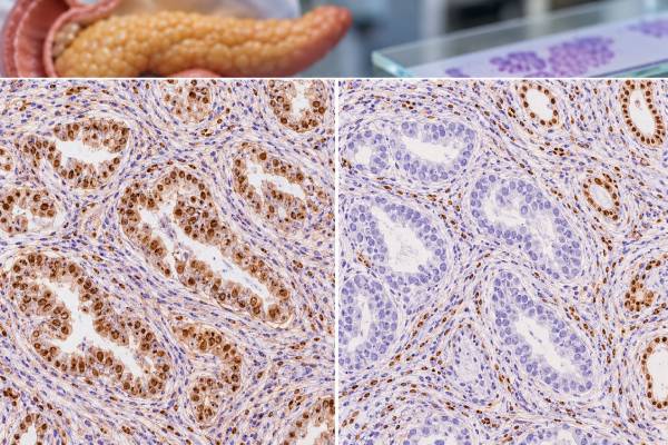
DPC4 immunohistochemistry is a tissue test that shows whether tumor cells retain or lose SMAD4 protein, a tumor-suppressor protein frequently altered in pancreatic ductal adenocarcinoma. DPC4 and SMAD4 refer to the same pathway target: the historical gene name DPC4 stands for “deleted in pancreatic carcinoma 4,” while the modern gene symbol is SMAD4. In practice, pathologists use SMAD4/DPC4 staining as one part of a larger diagnostic panel. Complete loss of staining in tumor cells, with preserved staining in nearby normal cells, supports an abnormal SMAD4 pathway and can be useful when pancreatic cancer is suspected. However, SMAD4 loss is not present in every pancreatic cancer and can occur in other malignancies, so it cannot establish tumor origin by itself. The result is most useful when combined with morphology, cytokeratin patterns, other pancreatic markers, clinical imaging, and—when treatment or diagnostic uncertainty warrants it—molecular testing.
- Loss of DPC4/SMAD4 staining supports pancreatic ductal adenocarcinoma in the right context, but it is not specific enough to diagnose pancreatic origin alone.
- Retained SMAD4 does not rule out pancreatic cancer because a substantial proportion of pancreatic ductal adenocarcinomas preserve the protein.
- True loss requires an internal positive control, such as normal stromal, inflammatory, or benign epithelial cells that still stain.
- The stain is usually interpreted as retained versus lost, not by a blood-style numeric “normal range.”
- Molecular testing may be needed when results are discordant or treatment decisions depend on genomic findings.
Table of Contents
- What DPC4/SMAD4 IHC Measures
- Why SMAD4 Matters in Pancreatic Cancer
- How DPC4 IHC Is Interpreted
- DPC4 in Diagnosis and Tumor-Origin Workups
- Limitations and Common Pitfalls
- Prognosis and Treatment Relevance
- What to Ask About Your Result
What DPC4/SMAD4 IHC Measures
DPC4 IHC detects SMAD4 protein in formalin-fixed tumor tissue. SMAD4 is a central signaling protein in the transforming growth factor beta pathway, which helps regulate cell growth, differentiation, and other cellular functions. When both copies of the SMAD4 gene are inactivated or deleted, the tumor may stop producing detectable SMAD4 protein. Immunohistochemistry can reveal that loss indirectly.
The test is performed on a biopsy, fine-needle aspiration cell block, or resection specimen. The laboratory applies an antibody to a thin tissue section and uses a color-producing detection system. The pathologist compares staining in tumor cells with staining in non-neoplastic cells on the same slide.
That comparison is essential. A report of “SMAD4 loss” is only trustworthy when internal control cells remain positive. Stromal cells, lymphocytes, endothelial cells, and benign ducts commonly provide this control. If everything on the slide is negative, the stain may have failed technically and should not be called true tumor loss.
SMAD4 staining is normally nuclear and can also have cytoplasmic expression depending on the antibody and laboratory method. In routine pancreatic pathology, the key interpretation is generally whether tumor expression is retained or lost. Some laboratories describe heterogeneous or reduced patterns, but the clinically useful distinction is usually complete absence in tumor cells versus preserved expression.
There is no serum concentration, percentage threshold, or universal 0-to-3 score comparable with HER2. DPC4 IHC is a tissue-expression test. The report may simply state “SMAD4 retained” or “SMAD4 lost.”
The marker is often ordered as part of a broader tumor immunohistochemistry panel, particularly for an adenocarcinoma involving the pancreas, bile ducts, liver, peritoneum, lung, or another organ where the primary site is uncertain.
Why SMAD4 Matters in Pancreatic Cancer
Pancreatic ductal adenocarcinoma, often shortened to PDAC, is driven by a characteristic set of molecular alterations. KRAS activation is very common, while tumor-suppressor pathways involving CDKN2A, TP53, and SMAD4 are frequently disrupted during tumor progression. SMAD4 is inactivated in roughly half of PDACs in many published series, although exact percentages vary by cohort, specimen type, and testing method.
That frequency makes SMAD4 unusually useful in pancreatic pathology. If an adenocarcinoma has complete tumor-cell loss of SMAD4 with positive internal controls, the finding is compatible with pancreatic ductal adenocarcinoma and can strengthen a diagnosis already suggested by anatomy and morphology.
The historical name DPC4 came from its discovery as a gene commonly deleted in pancreatic carcinoma. Modern pathology reports more often use “SMAD4,” but laboratories and clinicians may still say DPC4. The terms are usually referring to the same immunohistochemical target.
SMAD4 is not a screening marker. It does not detect early pancreatic cancer in blood, predict that a healthy person will develop pancreatic cancer, or replace imaging. It is applied after tissue has been sampled. The stain may be particularly helpful in small biopsies where the tissue architecture is limited and in metastatic tumors where the organ of origin is not immediately obvious.
Pancreatic adenocarcinoma can also be evaluated with markers such as CK7, CK19, CA19-9, mucins, and other lineage-associated stains depending on the differential diagnosis. A related MUC5AC IHC result may support pancreatobiliary or gastric-type differentiation in selected cases, but none of these markers is perfectly specific.
How DPC4 IHC Is Interpreted
The interpretation depends on two questions: do the tumor cells stain, and do the surrounding non-tumor cells stain appropriately?
| Pattern | What it usually means | Key caution |
|---|---|---|
| Tumor negative; internal controls positive | True SMAD4 protein loss is supported | Loss supports but does not prove pancreatic origin |
| Tumor and internal controls positive | SMAD4 expression is retained | Pancreatic ductal adenocarcinoma is not excluded |
| Tumor and all controls negative | Result may be technically uninterpretable | Repeat staining or another block may be required |
| Patchy or heterogeneous tumor staining | May reflect biology, fixation, or sampling | Interpret with morphology and consider repeat/confirmatory testing |
Complete loss is the most straightforward abnormal pattern. The tumor glands or malignant cells are unstained while adjacent normal tissue remains clearly positive. This pattern can correlate with biallelic SMAD4 inactivation, but IHC is still a protein-level surrogate rather than direct sequencing.
Retained staining means detectable SMAD4 protein remains in tumor cells. It does not necessarily mean the SMAD4 gene is completely normal. Some mutations may not abolish protein expression, and pancreatic cancers with intact SMAD4 can still have other major driver alterations.
Heterogeneous staining deserves careful review. A small biopsy may include only part of a heterogeneous tumor, and fixation can create edge effects or uneven staining. If the interpretation is clinically important, a pathologist may test another block or compare with molecular results.
A common misunderstanding is to equate “loss” with a higher tumor stage. SMAD4 loss is a molecular/pathway feature, not a TNM stage measurement. Stage is based on anatomic extent—tumor size or local invasion, lymph nodes, and distant metastases—not on this stain.
DPC4 in Diagnosis and Tumor-Origin Workups
One of the most useful applications of DPC4 IHC is distinguishing a suspected pancreatic primary from competing adenocarcinomas. For example, a patient may have a liver lesion with gland-forming carcinoma and a pancreatic mass on imaging. Loss of SMAD4 in the tumor can support the pancreatic lesion as the source, especially when the morphology and remaining immunophenotype also fit.
However, SMAD4 loss is not exclusive to PDAC. Abnormal SMAD4 can be found in other gastrointestinal and extra-gastrointestinal cancers. Therefore, a pathologist should not label an unknown metastatic adenocarcinoma “pancreatic” on SMAD4 loss alone.
Tumor-origin workups often use panels designed around realistic alternatives. Examples include:
- CK7 and CK20 to help describe epithelial differentiation patterns;
- CDX2 and SATB2 when colorectal origin is considered;
- TTF-1 and Napsin A for a possible lung adenocarcinoma;
- PAX8 for renal, thyroid, or Müllerian origin;
- GATA3 for breast or urothelial differentiation;
- organ-specific clinical and radiologic findings.
A CK7 IHC staining pattern can be useful in a pancreatic-versus-colorectal differential, but again the pattern is not unique to one organ.
SMAD4 can also help separate malignant pancreatic epithelium from benign mimics in selected circumstances. The finding of true SMAD4 loss in atypical glands supports neoplasia because benign pancreatic ducts should retain the protein. Still, not every PDAC loses SMAD4, so retained staining cannot be used to declare an atypical gland benign.
In cytology and small core biopsies, tissue conservation matters. Pathologists often prioritize stains that answer the most important diagnostic questions while preserving material for molecular profiling. Because SMAD4 can provide both diagnostic and biologic information with one stain, it may be included when the specimen is limited.
Limitations and Common Pitfalls
The most important limitation is sensitivity. Because many pancreatic ductal adenocarcinomas retain SMAD4, a normal-looking stain does not exclude pancreatic cancer. The test is better viewed as a supportive abnormality when lost than as a universal marker that must be abnormal in every PDAC.
Specificity is also incomplete. SMAD4 alterations occur in colorectal, biliary, gastric, ovarian, and other carcinomas. The rate and pattern differ by tumor type, but the existence of these non-pancreatic losses prevents the stain from acting as a single-organ fingerprint.
Other pitfalls include:
- Missing internal controls. Without positive non-neoplastic cells, apparent loss can be a failed stain.
- Scant tumor. A few atypical cells may be difficult to distinguish from unstained debris or benign structures.
- Necrosis and treatment effect. Damaged tissue may show weak or absent antigen preservation.
- Heterogeneity. One block can differ from another, especially in larger or treated tumors.
- Over-reading reduced intensity. Weak staining is not automatically equivalent to complete loss.
- Confusing IHC with sequencing. Protein expression and DNA mutation status are correlated but not interchangeable.
Another practical issue is nomenclature. “DPC4 negative,” “SMAD4 loss,” and “loss of DPC4 expression” usually communicate the same idea, but report wording can vary. If a result is unclear, ask whether the pathologist means complete tumor-cell loss with an intact internal control.
The stain also cannot distinguish pancreatic cancer from every pancreatobiliary cancer with certainty. Cholangiocarcinoma and ampullary carcinomas may share morphology and immunophenotypic features. Clinical location, imaging, endoscopic findings, and other stains remain essential.
Prognosis and Treatment Relevance
SMAD4 has been studied extensively as a prognostic biomarker in pancreatic cancer. Some studies associate SMAD4 loss with more aggressive biology, particular metastatic patterns, or poorer outcomes, while other datasets show weaker or context-dependent relationships. Because results vary by disease stage and treatment setting, SMAD4 IHC is not generally used by itself to assign prognosis for an individual patient.
It is more accurate to say that SMAD4 loss provides biologic context. It indicates disruption of an important tumor-suppressor pathway and may correlate with other genomic or immune features. Research continues into whether SMAD4 status can help predict treatment response or identify useful therapeutic vulnerabilities.
For routine treatment selection, established clinical factors remain more important: stage, resectability, performance status, prior therapy, germline genetics, and actionable tumor alterations. Modern pancreatic cancer workups may include broad molecular profiling to look for uncommon but clinically important findings such as BRCA1/2 or PALB2-related homologous recombination defects, mismatch-repair deficiency, NTRK or RET fusions, KRAS G12C, BRAF alterations, or other targets depending on current treatment options.
SMAD4 IHC does not replace those tests. A tumor can show SMAD4 loss yet still require comprehensive molecular profiling. Similarly, a retained stain does not imply that the tumor lacks actionable changes.
If the pathology report uses SMAD4 loss to support pancreatic origin, treatment decisions should still be made from the final integrated diagnosis. If the final diagnosis remains uncertain and that uncertainty would change therapy, additional molecular testing or expert pathology review may be appropriate.
What to Ask About Your Result
A useful discussion with your care team starts with the wording of the final pathology diagnosis rather than the stain in isolation. Consider asking:
- Was SMAD4/DPC4 retained or completely lost in the tumor?
- Were internal positive controls present, confirming that the stain worked?
- Does the overall panel favor pancreatic ductal adenocarcinoma, or is another primary site still possible?
- Which other IHC markers were used to establish tumor origin?
- Does the specimen contain enough tumor for molecular testing?
- Has germline genetic testing or tumor sequencing been recommended?
- Is SMAD4 status being used only diagnostically, or does the oncology team believe it changes prognosis or treatment in this specific case?
If you are reading a report that says “loss of SMAD4 expression,” the practical translation is that the tumor cells failed to show the protein while control cells retained it. That finding is common enough in pancreatic ductal adenocarcinoma to be useful, but not common enough—or specific enough—to stand alone. The pathologist’s final integrated interpretation carries more weight than the isolated marker.
For patients with a pancreatic mass, pathology is only one part of staging and treatment planning. Imaging, resectability assessment, tumor markers such as CA 19-9, clinical condition, and multidisciplinary review all contribute. The CA 19-9 blood test has a very different role from SMAD4 IHC: it is a circulating marker used mainly in clinical context and monitoring, whereas SMAD4 examines protein expression directly in tissue.
DPC4 can be especially useful when the specimen comes from a metastatic site rather than the pancreas. Complete tumor-cell loss with intact staining in surrounding non-neoplastic cells can support a pancreaticobiliary-type primary when the morphology and clinical findings point in that direction. But loss is not exclusive to pancreatic ductal adenocarcinoma. Other carcinomas can alter the SMAD pathway, so radiology, endoscopy, prior pathology, and a broader IHC panel remain necessary before assigning an organ of origin.
The specimen type also changes confidence. A resection usually provides enough tissue to compare multiple tumor areas and internal controls, whereas a fine-needle aspiration cell block or small core may contain only a few malignant glands. On a scant sample, retained staining is straightforward if tumor nuclei and cytoplasm are convincingly positive, but apparent loss deserves particular attention to controls. If all cells on the slide are weak or unstained, the assay may be technically noninformative rather than showing true biologic loss.
SMAD4 IHC should also be separated from inherited-risk testing. Loss of SMAD4 protein in a pancreatic cancer is usually a tumor finding and does not by itself establish an inherited cancer syndrome. Germline testing is a different analysis performed on blood, saliva, or another normal-tissue source and may be recommended based on pancreatic-cancer guidelines, age, family history, and treatment implications. Likewise, tumor sequencing can detect DNA alterations that IHC cannot fully characterize. These tests can complement one another, but a pathology phrase such as “SMAD4 loss” should not be read as a complete genetic report.
When an older pathology report uses “DPC4” and a newer report uses “SMAD4,” they are referring to the same tumor-suppressor protein in this IHC context. The naming difference does not represent two separate stains. If reports appear discordant, the more useful comparison is whether tumor-cell expression was retained or lost and whether adequate internal controls were documented.
If the report is being used to evaluate a suspected recurrence, ask whether the staining was performed on the new lesion or referenced from the original pancreatic tumor. A current biopsy can answer a different question from archived tissue, especially when the new mass has an uncertain origin.
References
- Characteristics of p53 and Smad4 immunohistochemistry in pancreatic ductal adenocarcinoma and validation by next-generation sequencing. 2026
- Histopathological and genetic landscape of pancreatic ductal adenocarcinoma and its variants. 2025 (Review)
- Pancreatic Cancer Biomarkers: Oncogenic Mutations, Tissue and Liquid Biopsies, and Radiomics-A Review 2023 (Review)
- Loss of SMAD4 Is Associated With Poor Tumor Immunogenicity and Reduced PD-L1 Expression in Pancreatic Cancer 2022
- The utility of SMAD4 as a diagnostic immunohistochemical marker for pancreatic adenocarcinoma, and its expression in other solid tumors. 2007
Disclaimer
DPC4/SMAD4 IHC is an ancillary pathology test and cannot diagnose pancreatic cancer or determine tumor origin on its own. Interpretation depends on internal controls, tumor morphology, other stains, imaging, and sometimes molecular testing. Discuss the complete pathology report and its treatment implications with your pathologist, oncologist, or treating clinician.





