
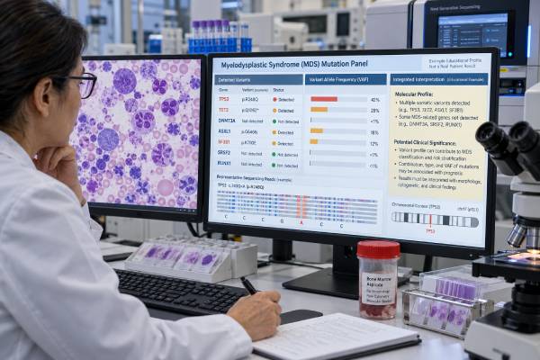
A myelodysplastic syndrome (MDS) mutation panel looks for acquired gene changes in blood-forming cells that can help define a myeloid clone, refine an MDS diagnosis, estimate prognosis, and guide treatment planning. Modern panels commonly use next-generation sequencing (NGS) to examine genes involved in RNA splicing, epigenetic regulation, transcription, signaling, and tumor suppression. Frequently altered genes include SF3B1, TET2, ASXL1, SRSF2, DNMT3A, RUNX1, U2AF1, STAG2, EZH2, and TP53, among others.
The result is not interpreted as a simple “MDS positive” or “MDS negative” test. Some mutations can occur in clonal hematopoiesis without MDS, while other molecular patterns strongly affect classification or risk. Diagnosis still integrates persistent cytopenias, bone marrow morphology, blast percentage, chromosome studies, and clinical history. Molecular findings have become especially important for the Molecular International Prognostic Scoring System (IPSS-M), which combines mutations with blood counts, blasts, and cytogenetics to provide a more individualized risk estimate than older clinical-cytogenetic models alone.
- What it measures: An MDS mutation panel detects somatic variants in genes recurrently altered in myeloid neoplasms, usually by NGS.
- What an abnormal result means: A pathogenic mutation can support clonality and may affect classification or prognosis, but most mutations do not diagnose MDS by themselves.
- High-risk findings: Multihit TP53 and mutations such as FLT3 or KMT2A partial tandem duplication can carry important adverse prognostic weight in molecular risk models.
- Potentially favorable context: SF3B1 mutation is linked to a distinct lower-risk MDS biology in appropriate cases, although co-mutations and other disease features can modify risk.
- Preparation: No fasting is usually needed; testing may use peripheral blood or bone marrow, but marrow is often obtained because morphology and blast assessment remain central to diagnosis.
Table of Contents
- What an MDS mutation panel tests
- When molecular testing is used
- Common MDS genes and what they can mean
- How mutation results affect MDS diagnosis and classification
- IPSS-M, prognosis, and risk categories
- How to read an MDS NGS report
- Limitations, follow-up, and next steps
What an MDS mutation panel tests
An MDS mutation panel usually examines dozens of genes that are recurrently altered in myeloid cancers. The exact gene list varies by laboratory. Unlike a single-gene assay, NGS can detect several mutations at the same time and report the molecular profile of the clone.
Most panels look for small DNA changes such as single-nucleotide variants and short insertions or deletions. Some assays also detect selected larger alterations, copy-number changes, or internal tandem duplications, but these capabilities are not universal. Cytogenetic testing and fluorescence in situ hybridization may still be needed because an NGS panel does not automatically capture every chromosome abnormality relevant to MDS.
The genes fall into biologic groups. Common examples include:
- RNA-splicing genes: SF3B1, SRSF2, U2AF1, and ZRSR2;
- epigenetic regulators: TET2, DNMT3A, ASXL1, EZH2, and IDH1/IDH2;
- transcription and differentiation genes: RUNX1, ETV6, and GATA2;
- cohesin-complex genes: STAG2 and related genes;
- signaling genes: NRAS, KRAS, CBL, JAK2, and FLT3; and
- tumor-suppressor genes: especially TP53.
These alterations can provide evidence that abnormal blood formation is clonal rather than purely reactive. However, clonality is not the same thing as MDS. Age-related clonal hematopoiesis can produce mutations in genes such as DNMT3A, TET2, and ASXL1 in people who do not meet diagnostic criteria for a myeloid neoplasm.
That is why an MDS panel is best viewed as one layer of an integrated workup. The molecular result becomes meaningful when combined with the blood count, smear, marrow findings, cytogenetics, and the reason the patient is being evaluated.
A broader hematologic cancer biomarker panel may include molecular, immunophenotypic, and cytogenetic methods, but an MDS-focused panel is designed specifically around recurrent myeloid mutations.
When molecular testing is used
Molecular testing is commonly ordered when unexplained cytopenias or abnormal marrow findings raise concern for MDS. Cytopenia means a low level of one or more blood-cell types, such as anemia, neutropenia, or thrombocytopenia. Before labeling the problem MDS, clinicians also look for reversible or non-neoplastic causes such as nutritional deficiencies, medications, alcohol exposure, infection, autoimmune disease, kidney or liver disease, and other marrow disorders.
An MDS mutation panel can be useful in several situations:
- persistent unexplained cytopenia with equivocal marrow morphology;
- a new bone marrow diagnosis of MDS for molecular characterization;
- risk assessment before choosing lower-intensity therapy, clinical-trial treatment, or allogeneic stem-cell transplantation;
- suspected therapy-related myeloid neoplasm after chemotherapy or radiation;
- follow-up of a known clone when the result could change management; or
- evaluation of progression when blood counts, blasts, or clinical features worsen.
A mutation result may help distinguish clonal cytopenia of undetermined significance (CCUS) from an unexplained cytopenia without a detectable clone. CCUS means a person has persistent cytopenia plus one or more myeloid-associated somatic mutations but does not yet satisfy full morphologic or genetic criteria for a defined myeloid neoplasm. CCUS carries a higher risk of progression than clonal hematopoiesis without cytopenia, but it is not automatically MDS.
Bone marrow remains important because MDS diagnosis depends heavily on dysplasia, cellularity, blast percentage, fibrosis, and other architectural findings. Molecular testing should complement—not replace—those observations.
If blasts rise or the molecular profile suggests evolution toward acute myeloid leukemia, additional AML-focused classification and monitoring may be needed. A leukemia flow cytometry panel can help characterize abnormal blasts when an acute leukemia is suspected.
Common MDS genes and what they can mean
No single list captures every clinically relevant MDS gene, and the meaning of a mutation depends on its exact variant, allele state, co-mutations, cytogenetic findings, and disease phenotype. Still, several genes recur often enough to have established diagnostic or prognostic importance.
| Gene or group | Typical role in interpretation | Important nuance |
|---|---|---|
| TP53 | Major adverse-risk marker, especially with multihit or biallelic inactivation | Allele state, VAF, cytogenetics, and genomic complexity matter |
| SF3B1 | Can define a distinct MDS subtype and often supports more favorable biology | Risk can worsen with certain co-mutations or higher-risk disease features |
| ASXL1 | Common myeloid mutation that can contribute adverse prognostic information | Also occurs in clonal hematopoiesis, so it is not diagnostic alone |
| RUNX1 | Associated with myeloid dysplasia and may increase molecular risk | Context matters because RUNX1 alterations occur across several myeloid neoplasms |
| SRSF2, U2AF1, ZRSR2 | Spliceosome mutations support a clonal myeloid process and influence phenotype/risk | Different genes and hotspot variants have different effects |
| TET2, DNMT3A | Frequent early clonal hematopoiesis mutations | May be present years before overt MDS and need careful clinical correlation |
| EZH2, STAG2, ETV6 | Can add adverse molecular information in prognostic models | Usually interpreted with the full mutation set rather than in isolation |
| FLT3, KMT2A-PTD | Relatively uncommon in MDS but strongly adverse in IPSS-M when present | May signal aggressive biology or disease evolution |
TP53: allele state matters
TP53 is one of the most important high-risk findings in MDS, but “TP53 positive” is not specific enough for modern interpretation. Some patients have one detectable TP53 alteration, while others have multihit or biallelic TP53 inactivation caused by multiple mutations, deletion of the other allele, or copy-neutral loss of heterozygosity.
Multihit TP53 disease is associated with particularly adverse outcomes and is recognized as a biologically distinctive group in contemporary classification systems. TP53 abnormalities also often occur with complex cytogenetic changes. A TP53 mutation result in blood cancer therefore needs interpretation beyond the gene name alone.
SF3B1: favorable does not mean harmless
SF3B1 mutation is strongly associated with an MDS phenotype involving ring sideroblasts and can define a molecularly characterized subtype under modern classifications when other criteria are met. In population-level studies, SF3B1-mutated MDS often has a more favorable natural history than many other molecular groups.
That does not mean every SF3B1-positive case is low risk. Excess blasts, adverse cytogenetics, transfusion dependence, or unfavorable co-mutations can outweigh part of the favorable signal. The entire clinical-molecular profile matters.
ASXL1, RUNX1, and spliceosome genes
ASXL1 and RUNX1 are commonly discussed as adverse or higher-risk molecular findings in myeloid disease, while SRSF2 and U2AF1 can also influence outcome and disease phenotype. These mutations are particularly useful when incorporated into a validated prognostic framework rather than treated as isolated labels.
For example, a RUNX1 mutation test may support a myeloid clone, but RUNX1 status must be interpreted with morphology, blasts, and other molecular findings.
How mutation results affect MDS diagnosis and classification
MDS remains an integrated diagnosis. A mutation panel can strengthen or refine the diagnosis, but the report must be read alongside the complete blood count, bone marrow aspirate and biopsy, chromosome analysis, and clinical history.
Modern WHO and International Consensus Classification systems give genetics a larger role than older schemes. Certain molecular abnormalities can help define disease categories, while others mainly support clonality or prognosis. The systems are not identical, so a pathology report may use terminology that depends on which classification framework the laboratory follows.
Three distinctions are especially important.
First, a mutation can be present without MDS. Clonal hematopoiesis of indeterminate potential (CHIP) is common with aging and is defined by a blood-cell clone carrying a somatic mutation in the absence of unexplained cytopenia or a diagnosed hematologic neoplasm. If cytopenia is present but MDS criteria are not met, the term CCUS may apply.
Second, some mutations have stronger disease-defining value than others. SF3B1 and TP53 are prominent examples in current MDS classification, although the exact required criteria differ by system. The interpretation may depend on blast percentage, variant allele frequency, allelic state, and coexisting cytogenetic abnormalities.
Third, molecular findings can reveal that a case belongs elsewhere in the myeloid spectrum. A patient initially suspected to have MDS may instead meet criteria for an MDS/MPN overlap neoplasm, AML, or another clonal disorder once the marrow, blood, cytogenetics, and genetics are integrated.
This is why a laboratory comment such as “mutation associated with myeloid neoplasms” should not be read as a final diagnosis. The most useful question is not simply whether a mutation is present, but whether the complete pattern explains the patient’s cytopenias and marrow findings.
IPSS-M, prognosis, and risk categories
The Molecular International Prognostic Scoring System, or IPSS-M, combines molecular data with the same broad clinical domains that have long mattered in MDS: blood counts, bone marrow blasts, and cytogenetic abnormalities. The model was developed from nearly 3,000 patients and externally validated in a separate cohort. It incorporates the mutational status of 31 genes and assigns patients to six risk categories.
In the original development study, at least one oncogenic genomic alteration was identified in about 94% of patients. TP53 multihit status, FLT3 mutation, and KMT2A partial tandem duplication were among the strongest adverse genetic predictors. SF3B1 was associated with more favorable outcomes, but its effect depended on the co-mutation pattern. Compared with the Revised International Prognostic Scoring System (IPSS-R), IPSS-M improved prediction of survival, leukemia-free survival, and leukemic transformation and reassigned a substantial fraction of patients to a different risk category.
What the six IPSS-M categories mean
IPSS-M produces a continuous score that maps into six groups, commonly described from very low through very high risk. The category estimates the expected behavior of MDS at a population level. Higher-risk groups have, on average, shorter survival and a greater chance of progression to AML.
The category is not a countdown or a guarantee for an individual patient. Age, fitness, other illnesses, transfusion burden, treatment response, transplant eligibility, and personal goals still influence decisions.
Why molecular risk can change treatment discussions
A person who looks lower risk by blood counts and cytogenetics may move into a higher IPSS-M category because of adverse mutations. The reverse can also occur. This can influence how strongly a team considers closer monitoring, disease-modifying therapy, a clinical trial, or referral for allogeneic stem-cell transplantation.
Molecular risk is especially useful because two patients with similar hemoglobin, platelets, neutrophils, and blast percentages can have very different clonal biology. IPSS-M captures part of that difference.
However, treatment should not be based on the IPSS-M number alone. The score is a prognostic tool, not a stand-alone treatment prescription.
How to read an MDS NGS report
An MDS molecular report typically lists the gene, exact DNA or protein variant, classification, and variant allele frequency (VAF). Some reports also state sequencing depth, assay sensitivity, coverage limitations, and whether copy-number or structural variants were assessed.
Pathogenic versus uncertain variants
A pathogenic or likely pathogenic variant has sufficient evidence to be clinically meaningful. A variant of uncertain significance (VUS) does not. A VUS should not be treated as proof of MDS or as an established high-risk mutation until additional evidence supports reclassification.
What VAF does—and does not—tell you
VAF is the percentage of sequencing reads that contain a variant. It gives a rough sense of the clone’s representation in the sampled DNA, but it is not the percentage of cancer cells. Each cell normally carries two copies of most genes, and copy-number changes, loss of heterozygosity, sample purity, and lineage distribution can alter the relationship between VAF and clone size.
A small VAF may reflect an early or minor clone. A higher VAF may reflect a larger clone or, for some genes, loss of the normal allele. The same numerical VAF therefore has different implications depending on the gene and genomic context.
One mutation versus several
MDS often evolves through sequential mutation acquisition. Multiple pathogenic variants may indicate a more complex clone, but the number of mutations is not itself a universal risk score. The identities and combinations matter. For example, SF3B1 with a relatively favorable background differs biologically from SF3B1 accompanied by strong adverse features.
The report should also be compared with prior molecular results if available. A new mutation, expanding clone, rising blast percentage, or worsening cytopenias can suggest clonal evolution, although treatment decisions should not be made from VAF movement alone.
Limitations, follow-up, and next steps
A normal or negative NGS panel does not rule out MDS. No panel captures every possible pathogenic alteration, and some cases are defined mainly by morphology or cytogenetics. Technical sensitivity also matters: a small clone can fall below the assay’s detection limit.
Conversely, a positive panel does not prove MDS. Mutations associated with myeloid neoplasms become more common with age and can occur in otherwise healthy people. The risk of overcalling MDS is greatest when a molecular result is separated from the marrow and clinical context.
After testing, useful questions include:
- Which variants are pathogenic or likely pathogenic, and which are VUS?
- What are the VAFs, and is TP53 allele state known if TP53 is mutated?
- Do the molecular findings satisfy or support a specific WHO or ICC disease category?
- What do the bone marrow morphology, blast percentage, and cytogenetics show?
- What is the IPSS-M category, and how does it compare with IPSS-R?
- Does the result change monitoring, therapy, clinical-trial eligibility, or transplant planning?
- Is repeat testing needed if blood counts or blasts change?
The most useful molecular profile is one that answers a clinical question. In an uncertain cytopenia, the question may be whether a clonal myeloid process is present. In established MDS, it may be how the mutations affect classification and prognosis. At progression, the question may be whether the clone has evolved toward a more aggressive state.
Molecular results can also overlap with age-related clonal hematopoiesis. A TET2 mutation, for example, may occur in MDS but can also be found in clonal hematopoiesis without overt cancer. That distinction is made from the whole clinical picture, not the gene name.
Prompt hematology review is warranted when cytopenias are severe or worsening, blasts increase, or symptoms suggest complications such as serious infection, symptomatic anemia, or significant bleeding. Molecular testing adds precision, but safe management still begins with the patient’s current blood counts and clinical condition.
References
- Molecular International Prognostic Scoring System for Myelodysplastic Syndromes. 2022
- Myelodysplastic syndromes: 2023 update on diagnosis, risk-stratification, and management 2023 (Review)
- Real-World Validation of Molecular International Prognostic Scoring System for Myelodysplastic Syndromes 2023
- Assessment and validation of the molecular international prognostic scoring system for myelodysplastic syndromes 2023
- Comparison of the 2022 world health organization classification and international consensus classification in myelodysplastic syndromes/neoplasms 2024
Disclaimer
This article is for general education and is not a diagnosis or treatment plan. MDS mutation results must be interpreted by a qualified hematology team with the complete blood count, bone marrow findings, cytogenetics, clinical history, and other tests. Seek prompt medical care for fever with severe neutropenia, significant bleeding, chest pain, severe shortness of breath, fainting, or other urgent symptoms.





