
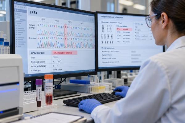
A TP53 mutation test looks for changes in one of the body’s most important tumor-suppressor genes. In blood cancers, TP53 abnormalities are especially important in myelodysplastic neoplasms (MDS), acute myeloid leukemia (AML), chronic lymphocytic leukemia (CLL), and several lymphomas because they can identify biologically high-risk disease and influence treatment planning. The test is usually performed by next-generation sequencing on blood or bone marrow and is often part of a larger cancer-gene panel. A positive result does not have one universal meaning: doctors consider the exact variant, variant allele frequency (VAF), whether the second TP53 copy is also lost or altered, chromosome findings, disease type, and treatment history. In AML and MDS, multi-hit or biallelic TP53 disruption is strongly linked to complex chromosomes, treatment resistance, short remissions, and high relapse risk. TP53 mutations can also appear in clonal hematopoiesis without overt leukemia, particularly after aging or prior cancer therapy. Accurate interpretation therefore requires the full hematology picture rather than the mutation alone.
- A pathogenic TP53 mutation is an important high-risk marker in many blood cancers, but its effect depends on the specific leukemia, lymphoma, or myeloid neoplasm.
- In AML and MDS, biallelic or multi-hit TP53 disease is particularly adverse and is often associated with complex karyotypes and resistance to standard therapy.
- A TP53 mutation is not the same as del(17p); sequencing detects gene variants, while cytogenetic/FISH testing detects loss of chromosome material, and both may be needed.
- Variant allele frequency helps describe the clone but does not by itself prove biallelic disease or measure exact tumor percentage.
- Some TP53 variants in blood can reflect clonal hematopoiesis rather than overt cancer, so the result must be matched to blood counts, marrow findings, and other molecular abnormalities.
Table of Contents
- What TP53 Does and How It Is Tested
- What a Positive TP53 Mutation Means
- TP53 in AML and MDS
- Single-Hit vs Multi-Hit TP53
- Variant Allele Frequency and Clonal Hematopoiesis
- Prognosis and Treatment Implications
- What to Do After a TP53 Result
What TP53 Does and How It Is Tested
TP53 encodes the p53 protein, a cellular stress-response regulator sometimes called the “guardian of the genome.” When DNA is damaged or a cell is under abnormal growth pressure, normal p53 can pause the cell cycle, support DNA repair, or trigger cell death. Loss of this safeguard allows genetically unstable cells to survive and accumulate additional abnormalities.
Most hematology laboratories test TP53 with next-generation sequencing (NGS), often as part of a multigene panel. Blood can be used when circulating abnormal cells are present; bone marrow is common in suspected or established myeloid disease. Some assays sequence the coding regions and nearby splice sites, while broader platforms can also assess copy-number changes.
A typical molecular report may list:
- the exact TP53 variant;
- whether it is classified as pathogenic, likely pathogenic, or a variant of uncertain significance;
- the VAF;
- assay sensitivity and coverage; and
- other mutations detected on the panel.
Sequencing and chromosome testing answer different questions. TP53 sits on chromosome 17p13.1. A deletion of 17p can remove one TP53 copy, but a patient may have a TP53 mutation without del(17p), del(17p) without a detected coding mutation, or both. Conventional karyotyping, FISH, chromosomal microarray, copy-number analysis, and sequencing can therefore be complementary.
The broader hematologic cancer biomarker panel matters because TP53 rarely acts in isolation. The diagnosis and risk category depend on the disease, chromosome pattern, co-mutations, blast percentage, morphology, and clinical history.
A VUS should not be treated like a confirmed pathogenic TP53 mutation. Variant classification can change as evidence develops, so laboratories may update interpretation over time.
What a Positive TP53 Mutation Means
A positive pathogenic TP53 result means that a detectable population of tested cells carries an alteration expected to impair normal p53 function or otherwise contribute to cancer biology. It does not automatically tell whether the person has leukemia, how advanced the disease is, or how long they will live.
The clinical meaning depends first on what disease is being evaluated. In AML and MDS, TP53 mutations define a particularly high-risk biologic group, especially when both TP53 alleles are affected. In CLL, TP53 mutation and del(17p) are major predictive markers because they influence treatment selection. In lymphoma, TP53 abnormalities can also mark aggressive biology, although the strength and therapeutic implications vary by subtype.
The same mutation may occasionally be found outside overt cancer. TP53-mutated clonal hematopoiesis can develop with age and is particularly selected by prior exposure to cytotoxic chemotherapy or radiation. In a person with normal or only mildly abnormal counts and no diagnostic marrow findings, a TP53 clone may represent clonal hematopoiesis rather than leukemia. Because TP53 is a higher-risk clonal hematopoiesis gene than some common age-related drivers, the finding usually deserves careful hematologic context and follow-up.
A positive result should therefore trigger four questions:
- Is the variant truly pathogenic?
- What hematologic diagnosis is present, if any?
- Is TP53 affected on one allele or both?
- What other molecular and cytogenetic abnormalities are present?
Those questions turn “TP53 positive” from a broad warning label into a clinically useful result.
TP53 in AML and MDS
TP53 mutations occur in roughly 8%–12% of MDS and AML overall, with higher rates in therapy-related disease, complex karyotypes, and older patients. These cases often have multiple chromosome losses and structural abnormalities, reflecting profound genomic instability.
In AML, current risk systems treat TP53-mutated disease as adverse-risk in the appropriate setting. The mutation is associated with lower response rates to conventional intensive chemotherapy, shorter remission duration, and high relapse rates. Even when a remission is achieved, the disease may return because resistant TP53-mutated clones persist.
In MDS, TP53 status is also highly important. Multi-hit TP53 MDS behaves much more aggressively than many lower-risk MDS categories and can progress rapidly. Modern classification systems recognize TP53-mutated myeloid neoplasia as a distinct or specially defined biologic group, although the World Health Organization and International Consensus Classification use somewhat different thresholds and naming rules.
TP53 does not replace the rest of the workup. AML and MDS evaluation still requires complete blood counts, marrow morphology, blast percentage, conventional cytogenetics, and a broader MDS mutation panel or myeloid NGS panel. Disease history also matters because prior chemotherapy or radiation can select TP53-mutant hematopoietic clones and contribute to therapy-related myeloid neoplasms.
Unlike mutations such as NPM1 that can be useful for sensitive leukemia-specific measurable residual disease testing, TP53 can persist in preleukemic or clonal hematopoiesis compartments. A persistent low-level TP53 variant after treatment may still be clinically concerning, but it should not automatically be interpreted exactly like a validated leukemia-specific MRD marker.
Single-Hit vs Multi-Hit TP53
The number of functional TP53 copies matters. Human cells normally have two alleles, one inherited from each parent. “Single-hit” generally means one allele is altered while the other appears intact. “Multi-hit,” “biallelic,” or “double-hit” refers to situations in which both functional copies are lost or disabled.
Biallelic disruption can occur in several ways:
- one TP53 mutation plus deletion of the other 17p allele;
- two separate pathogenic TP53 mutations;
- a mutation plus copy-neutral loss of heterozygosity, where the normal copy is lost and the mutant copy is duplicated; or
- other copy-number patterns that effectively remove normal TP53 function.
This distinction matters because multi-hit TP53 myeloid neoplasms generally have the strongest association with complex karyotype, treatment resistance, and poor survival. A single pathogenic mutation at a lower VAF may carry a different prognosis, especially if there is no evidence that the second allele is lost.
A sequencing report alone may not always establish allelic state. VAF provides clues, but copy-number and cytogenetic information are often needed. A very high VAF can suggest loss of the normal allele or a large clone, but it is not definitive proof. Likewise, two mutations can occur in the same clone or in separate subclones, which can require more detailed analysis to resolve.
This is why clinicians and pathologists may use the phrase “TP53 mutation” differently from “multi-hit TP53.” The latter is a more specific biologic concept with stronger prognostic implications in myeloid disease.
Variant Allele Frequency and Clonal Hematopoiesis
VAF is the percentage of sequencing reads at a genomic position that contain the variant. A TP53 VAF of 25% does not mean that exactly 25% of marrow cells are leukemia cells. In a simple diploid situation with a heterozygous mutation, a 25% VAF might roughly correspond to about half the cells carrying the mutation, but copy-number changes, aneuploidy, sample purity, normal-cell contamination, and subclones can change that relationship.
VAF is useful in three main ways. First, it helps estimate the relative size of a clone. Second, it can support interpretation of allelic state when combined with chromosome data. Third, serial VAF can show whether a clone is expanding or contracting, though small differences can reflect assay variation.
Low VAF does not make a TP53 mutation irrelevant. Sensitive NGS can identify small TP53-mutant clones, and some may expand under treatment pressure. This is particularly important after cytotoxic chemotherapy, where TP53-mutated hematopoietic stem cells can have a survival advantage.
Clonal hematopoiesis creates a key diagnostic caution. A person can have a somatic TP53 mutation in blood without meeting criteria for leukemia or MDS. The distinction depends on persistent cytopenias, marrow dysplasia, blast count, cytogenetics, additional mutations, and clinical findings. A clonal hematopoiesis mutation such as TET2 is more common in age-related CHIP, but TP53 and PPM1D are particularly associated with selection after genotoxic therapy.
High or near-50% VAF can also raise the possibility of a germline TP53 variant, especially with a suggestive personal or family cancer history. Blood is not an ideal tissue for confirming germline status because hematopoietic clones can mimic inherited variants. Confirmatory germline testing should use an appropriate non-blood source and genetics counseling when indicated.
Prognosis and Treatment Implications
In AML and MDS, TP53 mutation—particularly multi-hit disease—is among the strongest adverse molecular features. Standard regimens can produce responses, but remissions are often short. Hypomethylating agents, intensive chemotherapy, and venetoclax-containing combinations have all been used, yet none reliably overcomes the adverse biology.
Allogeneic stem cell transplantation remains the only potentially curative strategy for some patients with TP53-mutated myeloid disease, but relapse after transplant is common. Whether transplantation is appropriate depends on age, fitness, disease response, donor options, competing health risks, and patient preferences. Achieving the deepest possible remission before transplant may improve the chance of durable benefit, but outcomes remain less favorable than for many TP53-wild-type diseases.
Several TP53-directed or immune-based approaches have been investigated, including agents designed to restore mutant p53 function, macrophage checkpoint therapies, and other targeted combinations. Some promising programs have not produced durable phase 3 success, underscoring how difficult this biology is. Clinical trials are therefore particularly important for eligible patients.
A TP53 result should not be used to predict an exact survival time for an individual. Published outcomes are population averages and vary with the disease category, allelic state, cytogenetics, age, treatment era, transplant eligibility, and depth of response. New therapies are also changing benchmarks.
For CLL, the treatment implications are different enough to merit disease-specific interpretation. A CLL FISH panel and TP53 sequencing are used together because del(17p) and TP53 mutation can occur separately. Targeted therapies are generally preferred over traditional chemoimmunotherapy for TP53-aberrant CLL.
The key principle is that TP53 is both a risk marker and, in some diseases, a predictive marker—but the appropriate clinical action depends on the hematologic diagnosis.
What to Do After a TP53 Result
A new TP53 result should be reviewed with the complete pathology and laboratory report. If cancer is already diagnosed, the treating hematologist will usually incorporate it into formal risk classification and treatment planning. If the mutation was discovered incidentally, the first task is to determine whether there is evidence of an underlying hematologic neoplasm or clonal cytopenia.
Useful follow-up questions include:
- Is the variant pathogenic or only a VUS?
- What is the VAF and assay detection limit?
- Is there del(17p), loss of heterozygosity, or another TP53 mutation?
- Does the chromosome study show a complex karyotype?
- Which other mutations are present?
- Are blood counts persistently abnormal?
- Does bone marrow morphology establish AML, MDS, or another disease?
- Could prior chemotherapy or radiation have selected this clone?
- Does the personal or family history justify germline evaluation?
If TP53-mutated AML or MDS is diagnosed, referral to a center experienced in high-risk myeloid disease can be useful, especially when transplantation or clinical trials are being considered. Treatment decisions often require balancing the chance of response against toxicity and the risk of early relapse.
If the finding is clonal hematopoiesis without cancer, management may involve periodic blood counts and attention to changes rather than immediate anti-cancer therapy. Worsening anemia, neutropenia, thrombocytopenia, new circulating blasts, recurrent infections, unusual bleeding, or unexplained constitutional symptoms should prompt reassessment.
The most accurate way to understand TP53 is therefore not “positive equals hopeless.” It is a high-value molecular signal that must be placed in the right disease, allelic, cytogenetic, and treatment context. That integrated interpretation gives the result its real prognostic and clinical meaning.
TP53 testing can be especially complicated after prior chemotherapy because the mutation may predate overt leukemia. A small TP53-mutated hematopoietic clone can survive genotoxic treatment better than normal stem cells and expand years later. If a patient subsequently develops cytopenias, the clinician must distinguish therapy-associated clonal hematopoiesis from an actual therapy-related myeloid neoplasm using marrow morphology, blasts, cytogenetics, and additional mutations.
The chromosome pattern can add major context. TP53-mutated AML/MDS frequently has complex karyotype, including multiple losses and structural abnormalities. This combination generally signals more adverse biology than an isolated low-level TP53 mutation in an otherwise favorable molecular setting. For this reason, sequencing should be reviewed alongside the full karyotype rather than in a separate silo.
Response assessment also needs nuance. A falling TP53 VAF during therapy can support molecular response, but residual TP53 may reflect a preleukemic clone, and technical sensitivity differs between assays. Clinicians may combine molecular trends with marrow morphology, flow cytometry, chromosome studies, and other disease-specific MRD markers instead of using one TP53 percentage as the sole measure of remission.
The timing of testing matters as well. TP53 status should be established at diagnosis of a myeloid neoplasm and may be reassessed when disease changes, after therapy, or before transplantation if the result will affect risk interpretation. Clonal evolution can alter VAF and copy-number state, so a years-old molecular report may not describe the current disease.
A second pathology or molecular review can be valuable when the result will change a major decision and the allelic state is uncertain. Reconciling sequencing, FISH, karyotype, and copy-number data can clarify whether the case truly meets a multi-hit TP53 definition under the classification system being used.
References
- Diagnosis and management of AML in adults: 2022 recommendations from an international expert panel on behalf of the ELN 2022 (Guideline)
- The 5th edition of the World Health Organization Classification of Haematolymphoid Tumours: Myeloid and Histiocytic/Dendritic Neoplasms 2022 (Classification)
- TP53 in MDS and AML: Biological and clinical advances 2024 (Review)
- TP53 -Mutated Myeloid Neoplasms: 2024 Update on Diagnosis, Risk-Stratification, and Management 2025 (Review)
- TP53-Mutated Acute Myeloid Leukemia: Review of Treatment and Challenges 2025 (Review)
- TP53-mutated myelodysplastic syndromes and acute myeloid leukemia: a comprehensive overview of targeted approaches 2026 (Review)
Disclaimer
TP53 results can have major diagnostic and prognostic implications but cannot be interpreted safely without the underlying disease, allelic status, chromosome findings, and clinical context. A pathogenic blood TP53 variant can also represent clonal hematopoiesis or, less commonly, an inherited variant. Review the result with a hematologist and genetics professional when germline risk is a concern.





