
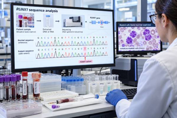
A RUNX1 mutation test looks for changes in the RUNX1 gene, a major regulator of blood-cell development. Acquired RUNX1 mutations occur in a subset of acute myeloid leukemia (AML), myelodysplastic neoplasms/syndromes (MDS), and related myeloid cancers. The result can help classify the disease, refine risk, explain a pattern of abnormal blood-cell production, and identify patients whose leukemia has features associated with myelodysplasia. A positive result does not, by itself, prove that someone has AML or MDS, and it does not predict one exact outcome. Interpretation depends on whether the variant is somatic or germline, the percentage of blasts, chromosome findings, other mutations, blood counts, prior blood disorders, and treatment response. RUNX1 also needs to be distinguished from RUNX1::RUNX1T1, a different genetic abnormality caused by the t(8;21) translocation. That fusion defines a separate AML subtype and generally has different prognostic implications from a point mutation or small insertion/deletion in RUNX1.
- A positive RUNX1 mutation means a pathogenic or likely pathogenic RUNX1 variant was detected in the tested cells, but diagnosis still requires the full blood and bone marrow picture.
- In AML, RUNX1 is one of the myelodysplasia-related genes used in modern molecular classification and risk assessment, although the exact category depends on other findings.
- RUNX1 mutations are often associated with higher-risk myeloid disease, but prognosis is not determined by RUNX1 alone. Co-mutations, cytogenetics, treatment, and disease stage matter.
- A blood or marrow RUNX1 mutation can occasionally be germline. Personal and family history may prompt confirmatory inherited-cancer testing using a non-blood tissue source.
- RUNX1 mutation is not the same as RUNX1::RUNX1T1 fusion. Reports should be read carefully because those abnormalities have different biological and clinical meanings.
Table of Contents
- What RUNX1 Does and What the Test Detects
- When RUNX1 Testing Is Used
- How RUNX1 Mutation Results Are Reported
- RUNX1 in AML and MDS Classification
- What RUNX1 Means for Prognosis
- Somatic Versus Germline RUNX1 Mutations
- Treatment, Follow-Up, and Common Pitfalls
What RUNX1 Does and What the Test Detects
RUNX1 is a transcription factor: a protein that helps turn groups of genes on or off. It is essential for normal hematopoiesis, the process by which bone marrow stem cells develop into red cells, white cells, and platelets. RUNX1 works with another protein, CBFB, to control differentiation and maturation of blood-cell precursors.
A mutation can reduce normal RUNX1 function or alter the way the protein controls gene expression. In a myeloid stem or progenitor cell, that can contribute to abnormal self-renewal, impaired differentiation, and the accumulation of additional cancer-driving changes.
Most clinical testing uses next-generation sequencing (NGS) as part of a myeloid gene panel. The assay examines RUNX1 along with other genes that commonly change in AML and MDS. Laboratories may detect single-nucleotide variants, small insertions/deletions, splice-site changes, and sometimes larger alterations depending on the platform.
RUNX1 is not one single “positive or negative” mutation. Different variants can affect different parts of the protein. A report therefore usually includes the exact DNA change, predicted protein change, pathogenicity classification, and variant allele frequency (VAF).
The VAF is the percentage of sequencing reads that contain the variant. It can provide clues about clone size, but it is not the same as the percentage of leukemia cells. For example, a VAF near 40% does not mean exactly 40% of marrow cells are malignant. Normal-cell contamination, copy-number changes, zygosity, and clonal structure all affect the number.
RUNX1 mutation testing may be included in a broader MDS mutation panel or AML molecular panel rather than ordered as a stand-alone assay.
When RUNX1 Testing Is Used
RUNX1 testing is most useful when clinicians are evaluating a suspected or confirmed myeloid neoplasm. Common reasons include persistent unexplained cytopenias, circulating blasts, abnormal marrow morphology, newly diagnosed AML, suspected MDS, or a myelodysplastic/myeloproliferative neoplasm.
The test may also be repeated when disease evolves, especially if a patient with MDS develops rising blasts or transforms to AML. Myeloid cancers change over time, and newly acquired variants can alter risk or clarify the biology of progression.
Typical specimens include:
- bone marrow aspirate, which is usually preferred at diagnosis because it directly samples the disease site;
- peripheral blood, which can be adequate when abnormal myeloid cells are circulating;
- non-blood tissue for germline confirmation, when an inherited RUNX1 variant is suspected.
RUNX1 results are interpreted with several other tests. A complete AML or MDS workup usually includes blood counts, morphology, bone marrow examination, chromosome analysis, FISH in selected settings, flow cytometry, and a multigene molecular panel. Other genes that often help define the same disease landscape include ASXL1, SRSF2, SF3B1, STAG2, BCOR, EZH2, U2AF1, ZRSR2, TP53, NPM1, FLT3, IDH1, and IDH2.
Testing for TET2 mutations and DNMT3A mutations is especially common on the same panel, although those variants can also appear in clonal hematopoiesis without an overt blood cancer.
No fasting is required for the gene test itself. If a bone marrow biopsy or aspiration is needed, preparation depends on the center and whether sedation is used.
How RUNX1 Mutation Results Are Reported
A molecular report may list RUNX1 as pathogenic, likely pathogenic, variant of uncertain significance (VUS), or not detected.
Pathogenic or likely pathogenic
This means the laboratory has enough evidence that the variant disrupts RUNX1 function or is associated with myeloid malignancy. In the proper disease context, the finding can contribute to classification and risk stratification.
The result still does not stand alone. A pathogenic RUNX1 variant found in a marrow with dysplasia and cytopenias has a different clinical context from the same molecular finding discovered incidentally in a blood sample from a person without cytopenias.
Variant of uncertain significance
A VUS is not the same as a disease-causing mutation. It means current evidence is insufficient to determine whether the change is harmful or benign. A VUS generally should not be used by itself to diagnose AML/MDS, assign an adverse-risk category, or make a transplant decision.
Not detected
A negative result means no reportable RUNX1 variant was found above the assay’s validated detection threshold. It does not exclude AML or MDS because most patients with these disorders do not have a RUNX1 mutation. Other molecular or chromosome abnormalities may explain the disease.
Why the VAF matters but does not give the whole answer
A higher VAF may suggest that the mutation is present in a larger clone. A value near 50% can also raise the question of a germline variant, especially when the same change is known to cause hereditary RUNX1 disorders. But VAF alone cannot prove germline status. Large somatic clones can also produce VAFs near 50%, and copy-number changes can distort the expected ratio.
RUNX1 in AML and MDS Classification
The meaning of RUNX1 changed significantly with the 2022 updates to AML classification.
Older World Health Organization systems included a provisional entity called AML with mutated RUNX1. That stand-alone category was removed from the 2022 WHO classification. Instead, RUNX1 is now interpreted as part of a broader molecular pattern of myelodysplasia-related disease.
The International Consensus Classification (ICC) and European LeukemiaNet (ELN) framework identify RUNX1 among the genes associated with myelodysplasia-related AML. The precise diagnostic and risk rules are not identical across systems, so a pathology report may use slightly different terminology depending on which classification the laboratory follows.
In practical terms, a RUNX1 mutation can support the idea that AML has a secondary-type or myelodysplasia-related genetic profile, particularly when it occurs with other mutations in genes such as ASXL1, SRSF2, STAG2, U2AF1, SF3B1, BCOR, EZH2, or ZRSR2.
This is different from the RUNX1::RUNX1T1 fusion created by t(8;21). That recurrent fusion defines core-binding-factor AML and is generally assigned to a favorable-risk group when other high-risk features do not dominate. A point mutation in RUNX1 should never be assumed to have the same meaning simply because the gene name appears in both reports.
RUNX1 can also be present in MDS. In that setting, it adds to the molecular profile but does not independently establish the diagnosis. MDS still requires integration of persistent cytopenias, marrow morphology, blast percentage, cytogenetics, and molecular findings. Newer prognostic systems such as molecular versions of the IPSS incorporate gene mutations to refine risk beyond traditional chromosome and blood-count variables.
What RUNX1 Means for Prognosis
RUNX1 mutations have historically been associated with lower remission rates and shorter survival in AML and MDS. However, current evidence shows that the effect is context-dependent rather than a fixed rule.
In AML, RUNX1 often travels with other adverse biological features: older age, antecedent MDS, secondary-type mutations, and unfavorable chromosome abnormalities. This makes it difficult to separate the independent effect of RUNX1 from the disease profile around it.
Meta-analyses have generally found poorer outcomes in RUNX1-mutated AML compared with RUNX1-wild-type AML, including lower complete remission rates and worse survival. Yet more recent real-world studies suggest that the mutation may lose some independent prognostic power once patients are grouped by modern genetic risk and treatment type.
For example, lower-intensity therapy combining a hypomethylating agent with venetoclax may produce outcomes that differ from historical chemotherapy cohorts. This does not make RUNX1 irrelevant; it means the significance of the mutation must be updated as treatment changes.
In MDS, RUNX1 is commonly associated with excess blasts, disease progression, and a higher probability of transformation to AML. But again, prognosis is driven by the complete genomic and clinical pattern, not a single gene.
A TP53 mutation, complex cytogenetics, multiple adverse mutations, high blast burden, and poor treatment response may have stronger or additive effects. Conversely, a patient with RUNX1 mutation but fewer competing adverse features may have a different outlook from someone with highly complex disease.
The most useful questions are therefore not “Is RUNX1 good or bad?” but:
- Which disease classification applies?
- What other mutations and chromosome abnormalities are present?
- Is the disease de novo, secondary, or therapy-related?
- What is the blast percentage?
- How did the disease respond to initial therapy?
- Is allogeneic stem cell transplantation appropriate for the overall risk profile?
Somatic Versus Germline RUNX1 Mutations
Most RUNX1 mutations found during AML or MDS testing are acquired somatic mutations in the abnormal blood-cell clone. A smaller but important group are inherited germline variants.
Germline RUNX1 pathogenic variants cause a hereditary condition often called RUNX1 familial platelet disorder with associated myeloid malignancy. People may have lifelong mild-to-moderate thrombocytopenia, abnormal platelet function, easy bruising or bleeding, and an increased lifetime risk of MDS, AML, and sometimes other hematologic malignancies.
Clues that may prompt germline evaluation include:
- low platelets or bleeding dating back to childhood or young adulthood;
- several relatives with thrombocytopenia, leukemia, MDS, or unexplained bleeding;
- a RUNX1 variant at a VAF suggestive of heterozygous germline status;
- the same RUNX1 variant persisting when the leukemia is in deep remission;
- a variant already known to cause hereditary RUNX1 disease.
Confirmatory germline testing should generally use DNA that is not derived from blood or marrow, because those tissues contain the abnormal hematopoietic clone. Cultured skin fibroblasts are often considered the most reliable specimen in a patient with a hematologic malignancy. Some centers may use other tissues depending on the clinical situation and laboratory policy.
Recognizing a germline RUNX1 variant matters for the patient and family. It can influence transplant donor selection, because a related donor carrying the same pathogenic variant may be unsuitable. It also permits genetic counseling and targeted testing of relatives who choose to be evaluated.
Treatment, Follow-Up, and Common Pitfalls
There is currently no standard drug prescribed solely because a leukemia has a RUNX1 point mutation. Treatment is chosen according to the overall AML or MDS category, age, fitness, disease burden, cytogenetic and molecular risk, prior therapy, and transplant eligibility.
For AML, options may include intensive induction chemotherapy, lower-intensity venetoclax-based therapy, targeted drugs for actionable co-mutations, clinical trials, and allogeneic stem cell transplantation. If the same panel identifies FLT3, IDH1, or IDH2 abnormalities, those findings may create specific targeted-treatment options.
For MDS, management ranges from observation and supportive care to growth factors, hypomethylating agents, newer disease-modifying therapies for selected molecular subtypes, clinical trials, or transplant. A RUNX1 mutation may contribute to the decision to monitor more closely or to consider higher-risk treatment strategies, but it is not used in isolation.
Unlike NPM1, RUNX1 is generally not an ideal stand-alone MRD marker. Some RUNX1 variants can persist as part of preleukemic or clonal hematopoiesis-related populations, and the biology can be more complex than a leukemia-specific fusion or mutation. Follow-up usually relies on marrow assessment, flow cytometry, cytogenetics, and molecular testing interpreted together.
Common mistakes include:
- confusing RUNX1 mutation with RUNX1::RUNX1T1 fusion;
- treating a VUS as if it were pathogenic;
- assuming a 50% VAF proves an inherited mutation;
- assuming a positive mutation alone diagnoses leukemia;
- interpreting the same mutation as equally prognostic in every treatment era;
- ignoring co-mutations and chromosome findings.
A useful follow-up discussion with the hematology team should cover the exact variant, disease classification, co-mutations, whether germline evaluation is appropriate, and whether the molecular result changes transplant planning. If the mutation was detected during an evaluation for unexplained cytopenias rather than established cancer, the next step may be careful longitudinal monitoring rather than immediate cancer treatment.
RUNX1 results also need careful distinction between somatic and germline disease. Most RUNX1 mutations found in AML or MDS are acquired in the malignant clone, but inherited pathogenic RUNX1 variants can cause familial platelet disorder with a predisposition to myeloid malignancy. A lifelong history of low platelets, unusual bleeding, or several relatives with thrombocytopenia, MDS, or leukemia can justify genetic counseling and confirmatory germline testing from a non-blood tissue source. Blood or marrow alone cannot reliably make that distinction once a clonal hematologic disorder is present.
For follow-up, RUNX1 is not always the best MRD marker. A mutation can be part of an early founder clone and may persist in preleukemic hematopoiesis, while other leukemia-defining alterations disappear. Conversely, a RUNX1 mutation can be lost or replaced during clonal evolution. MRD strategy should therefore use the most disease-specific validated marker available and may combine flow cytometry with molecular testing rather than tracking RUNX1 alone.
When reviewing a report, ask whether the mutation is pathogenic, what its VAF is, whether there are additional myelodysplasia-related mutations, and how the finding changes the formal WHO/ICC diagnosis or ELN risk category. Those details matter more than the gene name in isolation.
RUNX1 can also appear in clonal hematopoiesis, although it is less common than DNMT3A or TET2 in otherwise healthy people. A low-level RUNX1 clone found without diagnostic marrow abnormalities should therefore be interpreted with blood counts and co-mutations. Persistent cytopenias, multiple mutations, or an expanding clone increase the need for hematology follow-up.
References
- Diagnosis and management of AML in adults: 2022 recommendations from an international expert panel on behalf of the ELN 2022 (Guideline)
- The 5th edition of the World Health Organization Classification of Haematolymphoid Tumours: Myeloid and Histiocytic/Dendritic Neoplasms 2022 (Position Statement)
- Clinical Outcomes of Acute Myeloid Leukemia Patients Harboring the RUNX1 Mutation: Is It Still an Unfavorable Prognosis? A Cohort Study and Meta-Analysis 2022 (Systematic Review)
- AML classification in the year 2023: How to avoid a Babylonian confusion of languages 2023
- Investigations of the prognostic value of RUNX1 mutation in acute myeloid leukemia patients: Data from a real-world study 2024
- Potential factors underlying the progression of RUNX1-mutated MDS to AML 2025
Disclaimer
This article is for general education and does not replace interpretation by a hematologist, molecular pathologist, or genetic counselor. RUNX1 results must be interpreted with marrow findings, cytogenetics, co-mutations, treatment history, and the possibility of germline disease. Do not make treatment or family-testing decisions from a RUNX1 result without specialist review.





