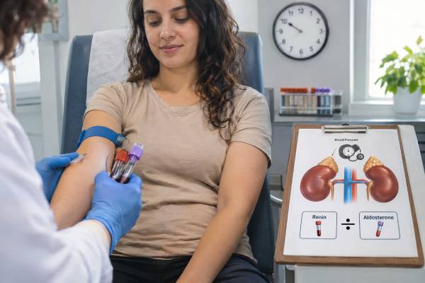
A renin blood test measures activity in the kidney-controlled system that regulates blood pressure, sodium, potassium, and fluid balance. Renin rises when the kidneys sense low blood flow, low sodium delivery, or sympathetic stimulation. It triggers angiotensin II and aldosterone, which help retain sodium and support blood pressure. Doctors rarely interpret renin alone. The result is usually paired with aldosterone and potassium, then considered in relation to posture, salt intake, collection time, kidney function, and blood-pressure medicines. Low renin with inappropriately high aldosterone can indicate primary aldosteronism. High renin with high aldosterone usually reflects a secondary response such as diuretic use, volume loss, or reduced kidney blood flow. Low renin with low aldosterone points toward other forms of mineralocorticoid-like hypertension or medication effects. The normal range depends on whether the laboratory measures plasma renin activity or direct renin concentration, so methods and units cannot be mixed.
- Renin is usually interpreted with aldosterone, potassium, blood pressure, and medication history.
- Low renin plus high or inappropriate aldosterone suggests primary aldosteronism.
- High renin commonly reflects diuretics, dehydration, ACE inhibitors, ARBs, renovascular disease, or heart failure.
- Plasma renin activity and direct renin concentration use different units and reference ranges.
- Posture, sodium intake, time of day, potassium, and many medicines can shift the result substantially.
Table of Contents
- What Renin Does
- Plasma Renin Activity Versus Direct Renin
- Why a Renin Test Is Ordered
- Preparation: Posture, Salt, and Medicines
- Causes of High Renin
- Causes of Low Renin
- Renin, Aldosterone, and ARR Patterns
- Follow-Up Testing and Treatment
What Renin Does
Renin is an enzyme released by juxtaglomerular cells in the kidneys. These cells respond to pressure in the kidney’s small arteries, sodium delivery to the macula densa, and sympathetic nerve signals. When effective circulating volume falls, renin secretion increases.
Renin cleaves angiotensinogen to angiotensin I. Angiotensin-converting enzyme then forms angiotensin II, which constricts blood vessels, supports blood pressure, stimulates thirst, and triggers aldosterone release from the adrenal cortex. Aldosterone increases sodium reabsorption and potassium excretion in the kidney.
As blood volume and pressure recover, renin should fall. This feedback means the absolute number is less informative than whether it is appropriate for the person’s blood pressure, sodium status, and aldosterone level.
A person with severe hypertension would ordinarily suppress renin. If renin remains high, the kidneys may be sensing reduced blood flow despite high systemic pressure, as in renal artery stenosis, or a medicine may be stimulating renin. A person with low blood pressure or dehydration would ordinarily raise renin; failure to do so can suggest medication effects, kidney damage, autonomic problems, or mineralocorticoid excess.
Renin is not an adrenal hormone, but it is central to adrenal aldosterone interpretation. The aldosterone blood test and renin together show whether the adrenal response is appropriate to kidney signaling.
Plasma Renin Activity Versus Direct Renin
Laboratories use two main approaches. Plasma renin activity, or PRA, measures how much angiotensin I is generated over time under standardized conditions. It is commonly reported in ng/mL/hour. Direct renin concentration, or DRC, measures the amount of renin protein and is often reported in mU/L or ng/L.
The values are correlated but not interchangeable. A PRA of 1 is not equivalent to a DRC of 1. The aldosterone-to-renin ratio changes numerically depending on the method. Clinical cutoffs must specify the renin assay and units.
PRA can be affected by substrate concentration and sample incubation. DRC is easier to automate but may be less precise at very low concentrations, where the denominator of the ARR becomes important. Modern laboratories use method-specific quality controls, yet variation remains.
The report should state:
- Renin method: PRA or DRC.
- Units.
- Collection posture.
- Time of collection when relevant.
- Reference interval.
- Aldosterone result and units if an ARR is calculated.
A result described only as “renin low” without these details cannot be compared reliably with another laboratory. Trends are easiest to interpret when the same method and collection conditions are used.
Some reports calculate the ARR automatically. Others provide the two analytes and leave calculation to the clinician. The ratio should never be interpreted without the actual aldosterone and renin values.
Why a Renin Test Is Ordered
The most common use is screening for primary aldosteronism in a person with hypertension. Low renin combined with aldosterone that is high or inappropriate produces an elevated ARR. Screening may be considered broadly in hypertension and is especially important with resistant hypertension, low potassium, an adrenal mass, sleep apnea, atrial fibrillation, or early-onset disease.
Renin is also measured when evaluating unexplained high or low potassium, low blood pressure, salt wasting, suspected adrenal insufficiency, renovascular hypertension, and certain inherited blood-pressure disorders.
Common clinical questions include:
- Is aldosterone elevated because renin is driving it, or is the adrenal gland acting autonomously?
- Is low potassium caused by aldosterone or another renal loss?
- Is a person with low blood pressure appropriately activating the renin system?
- Does primary adrenal insufficiency include mineralocorticoid deficiency?
- Is treatment with a mineralocorticoid receptor antagonist adequately blocking aldosterone effect?
- Could a monogenic low-renin hypertension syndrome be present?
In primary adrenal insufficiency, low aldosterone and sodium loss tend to raise renin. High renin can help show insufficient mineralocorticoid replacement in a treated patient, although symptoms, blood pressure, potassium, and sodium are also used.
Renin is not a stand-alone test for kidney artery narrowing. Many people with renovascular disease have variable results, and medicines strongly affect the value. Imaging is selected according to the clinical probability, kidney function, and whether intervention would be considered.
Preparation: Posture, Salt, and Medicines
Renin changes with posture. Standing shifts blood toward the legs and activates the renin system. A sample drawn after being upright is usually higher than one drawn supine. Laboratories may request morning collection after the patient has been awake and upright for at least two hours, followed by several minutes seated. Other protocols use a defined supine period.
Sodium intake has a strong effect. Low sodium raises renin; high sodium suppresses it. For primary aldosteronism screening, patients generally maintain their usual sodium intake and avoid deliberate restriction unless another medical condition requires it.
Potassium matters because low potassium can reduce aldosterone production and obscure primary aldosteronism. Potassium should be checked and corrected when safe before screening.
Medicines can change renin dramatically:
- Diuretics raise renin through sodium and volume loss.
- ACE inhibitors and ARBs raise renin by interrupting angiotensin signaling.
- Mineralocorticoid receptor antagonists and amiloride raise renin and alter potassium.
- Beta-blockers and central sympatholytics suppress renin.
- NSAIDs can suppress renin.
- Dihydropyridine calcium-channel blockers may raise renin or lower the ARR.
- Estrogen-containing contraceptives can alter direct renin measurements.
Do not stop blood-pressure medicine without supervision. Severe hypertension, heart failure, or kidney disease can make washout unsafe. The clinician may interpret on therapy, substitute less-interfering drugs, or repeat later.
Acute illness, dehydration, vomiting, diarrhea, strenuous exercise, and changes in kidney function also affect results. Record the collection time, posture, medicines, potassium, and recent salt or fluid changes.
Causes of High Renin
High renin usually means the kidneys perceive low effective circulation or the renin system is pharmacologically stimulated. Diuretics are a frequent cause. They increase sodium loss and reduce effective volume, which raises renin and often aldosterone.
ACE inhibitors and ARBs remove angiotensin II feedback, so renin can rise markedly. This is an expected drug effect, not evidence of a renin-secreting tumor.
Other causes include dehydration, vomiting, diarrhea, bleeding, low sodium intake, heart failure, cirrhosis, nephrotic syndrome, and renal artery stenosis. In heart failure or cirrhosis, total body fluid may be high while the kidneys still sense inadequate effective arterial volume.
Primary adrenal insufficiency can produce high renin because aldosterone is deficient and sodium is lost. The pattern includes low aldosterone, possible low sodium, high potassium, low blood pressure, and low cortisol. An adrenal insufficiency panel distinguishes this from a secondary aldosterone response.
Rare causes include renin-secreting kidney tumors, severe malignant hypertension, and inherited salt-wasting tubulopathies. These conditions have additional clinical and laboratory clues.
High renin is interpreted with aldosterone:
- High renin plus high aldosterone suggests a secondary response.
- High renin plus low aldosterone suggests impaired adrenal response, medication effect, or severe illness.
- High renin while taking a mineralocorticoid blocker may show effective blockade rather than disease progression.
A high result is not automatically treated. The underlying cause—volume depletion, medication, kidney perfusion, heart failure, or adrenal disease—determines management.
Causes of Low Renin
Low renin is common in hypertension, especially with older age, high sodium intake, chronic kidney disease, and certain ancestry-related physiologic patterns. It becomes diagnostically important when paired with aldosterone and potassium.
Primary aldosteronism is the leading endocrine consideration. Aldosterone-driven sodium retention suppresses renin. Aldosterone may be clearly high or merely inappropriate for the degree of renin suppression.
Beta-blockers, clonidine, methyldopa, NSAIDs, and high sodium intake can suppress renin. A high ARR caused by drug-suppressed renin may be a false positive if aldosterone is not elevated.
Low renin plus low aldosterone suggests a non-aldosterone mineralocorticoid effect. Possibilities include Liddle syndrome, apparent mineralocorticoid excess, licorice or glycyrrhizin exposure, Cushing syndrome, deoxycorticosterone excess, and some congenital adrenal enzyme disorders.
The DOC test may be considered when hypertension and low potassium occur with both renin and aldosterone suppressed. Monogenic causes are more likely with severe early-onset hypertension or a strong family history.
Low renin can also occur with reduced kidney renin-producing capacity. Diabetic kidney disease, chronic interstitial disease, and aging can blunt the response.
A low result should not be labeled primary aldosteronism unless aldosterone and the clinical setting support it. The denominator problem is important: dividing by a value near zero can make the ARR very large even when aldosterone is low.
Renin, Aldosterone, and ARR Patterns
The ARR is a screening tool that compares aldosterone with renin. A high ratio with suppressed renin and sufficiently high aldosterone supports primary aldosteronism. Cutoffs vary by method and unit.
| Renin | Aldosterone | Common interpretation |
|---|---|---|
| Low | High or inappropriate | Primary aldosteronism pattern |
| High | High | Secondary hyperaldosteronism |
| Low | Low | Low-renin hypertension from another mineralocorticoid or drug effect |
| High | Low | Adrenal impairment, ACE inhibitor/ARB effect, or severe illness |
Potassium adds severity information but is not a reliable screen by itself. Many people with primary aldosteronism have normal potassium. Low potassium may also result from diuretics, vomiting, diarrhea, or other kidney disorders.
The primary aldosteronism panel is more useful than renin alone because it integrates these relationships and identifies preparation issues.
Repeated testing may be needed when medicines or potassium distort the first result. A consistent suppressed renin despite an ACE inhibitor or ARB, which should raise renin, can be a meaningful clue.
Follow-Up Testing and Treatment
After a possible primary aldosteronism pattern, clinicians may repeat the ARR under improved conditions, perform a confirmatory suppression test, and assess candidacy for subtype testing. Adrenal CT shows anatomy, while adrenal vein sampling determines whether secretion is unilateral or bilateral in most surgical candidates.
High renin with suspected renovascular disease may lead to kidney imaging when the clinical setting supports it, such as abrupt hypertension, worsening kidney function after ACE inhibitor or ARB therapy, asymmetric kidney size, or recurrent flash pulmonary edema.
High renin with low aldosterone and suspected adrenal failure leads to cortisol, ACTH, electrolytes, and possibly ACTH stimulation. Low renin with low aldosterone and early severe hypertension may lead to genetic or steroid testing.
Treatment targets the cause. Primary aldosteronism may be treated with adrenal surgery or a mineralocorticoid receptor antagonist. Volume depletion is corrected carefully. Heart failure and cirrhosis need disease-specific therapy. Medication-related changes may be expected and beneficial.
Renin can be followed during mineralocorticoid replacement or blockade. In primary adrenal insufficiency, persistently high renin with low blood pressure and salt craving may suggest insufficient fludrocortisone, while low renin and hypertension may suggest excess. In primary aldosteronism treatment, a rise from suppressed renin may indicate adequate blockade, but potassium, kidney function, and blood pressure remain essential.
Questions after a result include:
- Was renin measured by PRA or DRC?
- What posture and sodium conditions were used?
- Which medicines affected the value?
- What were aldosterone and potassium at the same time?
- Is the renin response appropriate for my blood pressure and volume state?
- Does the pattern need repeat testing, suppression testing, imaging, or genetic evaluation?
Renin is best understood as a physiologic signal. Its value comes from showing how the kidneys, adrenal glands, medicines, and circulation are interacting.
Special situations that change renin interpretation
Pregnancy activates the renin-angiotensin-aldosterone system as blood volume expands. Renin and aldosterone can both rise substantially, so nonpregnant reference intervals and ARR cutoffs may mislead. At the same time, progesterone has mineralocorticoid-antagonist effects, and pregnancy-related hypertension introduces additional physiology. Suspected primary aldosteronism during pregnancy requires specialist interpretation and medication choices that protect both mother and fetus.
Age also changes the baseline. Renin tends to decline with aging, and low-renin hypertension becomes more common. A low value in an older adult may therefore be less specific than the same value in a teenager with severe hypertension. Young people with markedly suppressed renin, low aldosterone, and early family hypertension deserve consideration of monogenic disorders such as Liddle syndrome or apparent mineralocorticoid excess.
Chronic kidney disease can produce several patterns. Damage to renin-producing tissue may suppress renin, while renal ischemia, diuretics, fluid shifts, and heart failure can raise it. Reduced kidney function also increases the risk of potassium changes when RAAS-blocking drugs or mineralocorticoid antagonists are used. Interpretation should include estimated glomerular filtration rate, urine findings, medication history, and volume status.
Renovascular hypertension is more likely when blood pressure worsens abruptly, begins unusually early, accelerates after years of stability, or is accompanied by asymmetric kidneys, an abdominal bruit, recurrent flash pulmonary edema, or a notable creatinine rise after an ACE inhibitor or ARB. Renin may be high, but a normal value does not exclude renal artery disease. Imaging is pursued when the clinical probability is high enough and the result would affect management.
Renin specimen handling deserves attention. PRA samples may require rapid cooling or freezing to prevent continued angiotensin generation, while DRC has different stability requirements. Cryoactivation of prorenin can distort some measurements if samples are stored incorrectly. The collecting laboratory should follow the method’s protocol rather than treating renin like a routine chemistry test.
Serial renin values are most useful when conditions are comparable. A change after starting spironolactone, eplerenone, fludrocortisone, a diuretic, or an ACE inhibitor may reflect the expected drug action. Comparing values without documenting dose, posture, sodium intake, and potassium can create a false impression of disease progression.
In primary adrenal insufficiency, renin helps adjust mineralocorticoid replacement. The goal is not always a single exact number. Blood pressure lying and standing, salt craving, edema, sodium, potassium, and symptoms must agree. A very high renin with dizziness and salt craving may suggest under-replacement; hypertension, edema, low potassium, and suppressed renin may suggest excessive fludrocortisone.
In treated primary aldosteronism, renin can function as a response marker. Persistent suppression may mean that aldosterone effect remains inadequately blocked, sodium intake is high, or the medication dose is insufficient. A rise into a measurable range often accompanies effective blockade, but forcing renin upward at the cost of hyperkalemia, kidney injury, or symptomatic hypotension is not appropriate.
Licorice deserves specific mention. Natural licorice containing glycyrrhizin inhibits an enzyme that normally protects the mineralocorticoid receptor from cortisol. The resulting pattern can include hypertension, low potassium, metabolic alkalosis, low renin, and low aldosterone. Some chewing tobacco, herbal products, teas, and candies contain enough to matter. Stopping exposure can reverse the abnormality over time.
The timing of menstrual hormones and estrogen-containing products can affect some renin assays more than others. Direct renin concentration may be suppressed by estrogen-related changes, potentially inflating a DRC-based ARR. The laboratory method and contraceptive formulation should be considered rather than applying a blanket rule.
Renin should ultimately answer a physiologic question: are the kidneys appropriately responding to circulation, sodium, and aldosterone? A result interpreted in that framework is far more useful than labeling it high or low in isolation.
Blood pressure during collection adds context. A suppressed renin in a person with high blood pressure is physiologically different from suppressed renin during dehydration and low pressure. Orthostatic measurements can reveal volume depletion or autonomic dysfunction that a seated clinic value misses.
Aldosterone and renin may also be interpreted after a saline or captopril challenge. Those stimulated or suppressed values use protocol-specific cutoffs and should not be compared with a routine baseline range. The report should clearly identify whether the specimen was basal or part of a dynamic test.
People taking biotin or supplements should disclose them, although interference varies by assay. More important is a complete list of prescription and nonprescription blood-pressure products, including licorice, decongestants, NSAIDs, and herbal diuretics. These exposures can shift renin through real physiology even when they do not interfere analytically.
One abnormal renin value should not lead directly to adrenal or kidney imaging. The paired hormone pattern, repeated preparation, severity of hypertension, kidney function, and intended treatment determine whether imaging will answer a useful question.
If repeat testing is planned, the clinician should specify which conditions must stay the same and which should change. That comparison turns a second renin value into a controlled physiologic reassessment rather than another unrelated snapshot.
Laboratory flags should be read in context of the requested posture. A renin value that is normal after standing may be inappropriately low during volume depletion, while a value above a supine range may be expected after upright activity. The ordering question defines which comparison is meaningful.
References
- Primary Aldosteronism: An Endocrine Society Clinical Practice Guideline 2025 (Guideline)
- Primary aldosteronism 2.0: an update for clinicians on diagnosis and treatment 2023 (Review)
- The Spectrum of Dysregulated Aldosterone Production: An International Human Physiology Study 2024
- Subclinical Primary Aldosteronism and Cardiovascular Health: A Population-Based Cohort Study 2024
- Update on Low-Renin Hypertension: Current Understanding and Future Direction 2024 (Review)
Disclaimer
This article provides general education and cannot diagnose the cause of a high or low renin result. Renin depends on assay method, posture, sodium intake, potassium, kidney function, and medicines. Do not stop blood-pressure, heart, kidney, or steroid treatment without guidance from the prescribing clinician.