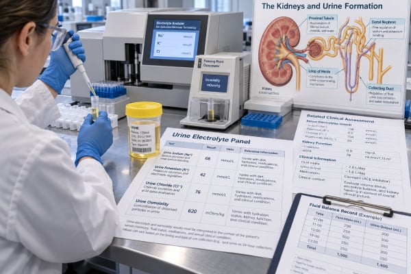
A urine electrolyte panel measures selected substances in urine—most often sodium, potassium, and chloride, with urine osmolality frequently added—to show how the kidneys are responding to a fluid, electrolyte, or acid-base problem. Unlike many blood tests, urine electrolytes usually do not have one “normal” value that applies in every situation. The correct result depends on what the kidneys should be doing at that moment. Low urine sodium can be appropriate when circulation is reduced and the kidneys are conserving salt, while the same low value may be unexpected after a large saline load. High urine potassium can be appropriate during hyperkalemia but inappropriate when blood potassium is already low. Urine osmolality shows whether the kidneys are concentrating or diluting water. Because medications, recent fluids, kidney disease, hormones, diet, and collection timing can change results quickly, a urine electrolyte panel is most useful when obtained with simultaneous blood electrolytes and interpreted as a physiologic pattern.
- Urine sodium and chloride mainly show renal salt handling, not total body sodium or chloride by themselves.
- Urine potassium helps distinguish kidney potassium loss from nonrenal loss when blood potassium is abnormal.
- Urine osmolality shows whether the kidneys are conserving water or producing dilute urine and is central to hyponatremia and polyuria evaluation.
- Spot urine values can change within hours after diuretics, IV fluids, vomiting, or changes in water intake, so timing matters.
- There is no universal “normal urine electrolyte panel.” The key question is whether each result is appropriate for the blood chemistry and clinical state.
Table of Contents
- What Is in a Urine Electrolyte Panel?
- How the Kidneys Handle Sodium, Potassium, and Chloride
- How to Interpret Common Urine Electrolyte Patterns
- Urine Osmolality and Water Balance
- Fractional Excretion and Derived Calculations
- Collection, Timing, Medications, and Pitfalls
- When Abnormal Results Need Follow-Up
- Practical Urine Electrolyte Panel Examples
What Is in a Urine Electrolyte Panel?
There is no single standardized urine electrolyte panel used by every laboratory. In practice, clinicians commonly order a combination of urine sodium, potassium, and chloride, and they often add urine osmolality. Creatinine, urea, calcium, magnesium, pH, or other measurements may be added depending on the clinical problem.
A panel can be collected from a random spot sample or from a timed collection. Spot samples are common in acute care because the goal is to understand what the kidneys are doing now. A 24-hour collection is more useful when the question concerns total daily excretion, such as sodium intake or kidney stone chemistry.
The four core measurements answer different questions:
| Measurement | Main physiologic question | Common uses |
|---|---|---|
| Urine sodium | Is the kidney retaining or excreting sodium? | Hyponatremia, AKI, volume assessment, salt balance |
| Urine potassium | Is the kidney appropriately conserving or excreting potassium? | Hypokalemia, hyperkalemia, aldosterone-related disorders |
| Urine chloride | Is chloride being conserved or lost? | Metabolic alkalosis, vomiting, diuretics, salt wasting |
| Urine osmolality | How concentrated is the urine? | Hyponatremia, hypernatremia, polyuria, water-balance disorders |
The panel is not a direct measure of kidney filtration. Filtration is assessed mainly with serum creatinine, cystatin C, and eGFR. Urine electrolytes instead evaluate tubular handling—how the nephron reabsorbs and secretes salt, potassium, acid-related ions, and water after filtration occurs.
This distinction matters because a person can have a nearly normal eGFR yet an important tubular or hormonal disorder that produces abnormal electrolyte handling.
How the Kidneys Handle Sodium, Potassium, and Chloride
The kidneys filter enormous amounts of sodium and chloride each day, then reabsorb almost all of it. Small changes in the fraction retained or excreted have large effects on extracellular fluid volume and blood pressure. Hormones such as the renin-angiotensin-aldosterone system, sympathetic nervous activity, and natriuretic peptides help control this process.
Sodium
When effective circulating volume falls, the kidneys usually conserve sodium, so urine sodium decreases. When the body has adequate or excessive circulating volume, sodium excretion generally rises. But this simple model can be disrupted by diuretics, adrenal disorders, kidney disease, recent IV fluids, and osmotic diuresis.
A urine sodium test is therefore interpreted against blood pressure, fluid status, medications, serum sodium, and kidney function.
Chloride
Chloride usually follows sodium, but the two can separate during acid-base disorders. In metabolic alkalosis with bicarbonate in urine, sodium may be excreted with bicarbonate while chloride stays low. This makes urine chloride particularly useful when vomiting, diuretics, or mineralocorticoid excess is being considered.
Potassium
Potassium handling is different because the distal nephron can actively secrete potassium. Aldosterone, sodium delivery to the distal nephron, urine flow, acid-base status, and blood potassium all influence how much potassium appears in urine.
When serum potassium is low, the expected kidney response is to reduce potassium excretion. Persistent urinary potassium loss despite hypokalemia suggests renal wasting from causes such as diuretics, mineralocorticoid excess, renal tubular disorders, or magnesium deficiency. When serum potassium is high, increased urinary potassium excretion is generally appropriate if kidney function and aldosterone action are intact.
How to Interpret Common Urine Electrolyte Patterns
Urine electrolytes are most useful as patterns rather than isolated numbers.
Hyponatremia
In true hypotonic hyponatremia, urine osmolality is often interpreted first. If urine is very dilute, usually below about 100 mOsm/kg, arginine vasopressin activity is largely suppressed and excess water intake or very low solute intake becomes more likely. If urine osmolality is above 100 mOsm/kg, the kidneys are retaining water under vasopressin influence, and urine sodium helps narrow the cause.
A low urine sodium often suggests reduced effective circulating volume, while a higher urine sodium can occur in syndrome of inappropriate antidiuresis, adrenal insufficiency, kidney salt wasting, or during diuretic treatment. Fixed cutoffs are imperfect because diet, CKD, and medications can alter them.
Hypokalemia
When blood potassium is low, clinicians ask whether potassium is leaving through the kidneys. A random urine potassium concentration alone is influenced by urine water content, so a potassium-to-creatinine ratio or 24-hour potassium excretion can be more informative. Acid-base status then helps organize the possibilities: diarrhea commonly causes metabolic acidosis with appropriate renal potassium conservation, while vomiting and diuretics often produce metabolic alkalosis.
Metabolic alkalosis
Urine chloride is the key panel component. A low value, often below 20 mmol/L, supports chloride depletion and a chloride-responsive process. A higher value during alkalosis points toward active diuretics, Bartter or Gitelman physiology, mineralocorticoid excess, or another renal chloride-wasting state.
Acute kidney injury
A low urine sodium or low fractional excretion of sodium can support a sodium-avid state in selected patients with acute kidney injury, but these findings are not diagnostic of “prerenal AKI” by themselves. Sepsis, glomerular disease, contrast exposure, CKD, and diuretics can produce exceptions. Urine sediment, hemodynamics, creatinine trend, and clinical context remain essential.
A broader acute kidney injury evaluation is therefore more reliable than deciding from a urine sodium value alone.
Urine Osmolality and Water Balance
Urine osmolality measures the number of dissolved particles per kilogram of urine water. It reflects the combined contribution of sodium, potassium, chloride, urea, and other osmoles. The kidney can vary urine osmolality over a very wide range, which is exactly why a fixed “normal” concentration is less useful than the physiologic response.
In an adult with normal kidney function, urine can become very dilute after excess water intake and very concentrated during water deprivation. Many laboratories report broad reference intervals, sometimes roughly 150–1,150 mOsm/kg for people older than infancy. Under ordinary diet and fluid intake, a midrange result is common, but any result must be matched to serum osmolality and the clinical state.
Important patterns include:
- Hyponatremia with urine osmolality below 100 mOsm/kg: appropriate water excretion; consider primary polydipsia or low-solute intake.
- Hyponatremia with urine osmolality >100 mOsm/kg: vasopressin is active; further evaluation with urine sodium and clinical context is needed.
- Polyuria with urine osmolality below 250–300 mOsm/kg: suggests water diuresis and raises the possibility of arginine vasopressin deficiency, resistance, or primary polydipsia.
- Polyuria with urine osmolality >300 mOsm/kg: osmotic or solute diuresis becomes more likely, such as glucosuria or high urea excretion.
An urine osmolality test is generally more reliable than urine specific gravity when large molecules such as glucose, protein, or radiographic contrast are present, because these substances disproportionately affect specific gravity.
Fractional Excretion and Derived Calculations
Urine electrolyte concentrations can be combined with blood values and urine creatinine to create derived indices. These calculations attempt to answer a more precise question than concentration alone.
Fractional excretion of sodium
The fractional excretion of sodium, or FENa, estimates what percentage of filtered sodium is excreted. A value below 1% has traditionally been associated with sodium avidity and prerenal physiology, while higher values can occur with tubular injury. A 2022 meta-analysis found good diagnostic performance in selected patients without CKD or diuretics, but accuracy fell substantially when those confounders were present.
This means FENa is useful when the clinical setting fits the assumptions, not as an automatic label for the cause of AKI.
Fractional excretion of urea
Fractional excretion of urea, or FEUrea, is sometimes used when diuretics make sodium indices harder to interpret. However, contemporary evidence shows that FEUrea also has only moderate diagnostic accuracy and is not consistently superior to FENa in diuretic-treated patients. It should be one data point among many.
Potassium indices
Spot urine potassium-to-creatinine ratio and 24-hour potassium excretion can help identify renal potassium wasting. The transtubular potassium gradient was historically used to infer distal potassium secretion, but it relies on assumptions about urine osmolality and collecting-duct physiology that are not always valid. Many clinicians now prefer simpler potassium excretion measures combined with serum potassium and acid-base status.
Urine anion and osmolal gaps
Urine sodium, potassium, chloride, and osmolality can also be used to estimate ammonium excretion during normal-anion-gap metabolic acidosis. These calculations are indirect and have important limitations; direct urine ammonium measurement is preferable when available.
Collection, Timing, Medications, and Pitfalls
A spot urine electrolyte sample is easy to collect, but interpretation can be difficult because kidney responses change quickly. Record what happened before the sample whenever possible: recent vomiting or diarrhea, IV saline, diuretics, food intake, water intake, blood pressure changes, and urine output.
Diuretics are particularly important. Loop and thiazide diuretics raise urine sodium and chloride while active and can increase potassium loss. A urine sample collected soon after a dose may therefore look very different from one collected the next morning. Mineralocorticoid antagonists, sodium-glucose cotransporter-2 inhibitors, acetazolamide, and other medications also influence urine chemistry.
Common mistakes include:
- interpreting urine sodium without confirming serum osmolality in hyponatremia;
- calling a low urine sodium “dehydration” when heart failure or cirrhosis could produce low effective arterial volume despite fluid overload;
- using urine potassium concentration without considering urine dilution;
- assuming a high urine chloride always means excess salt intake;
- applying FENa cutoffs despite diuretics or chronic kidney disease;
- comparing a spot concentration with a 24-hour reference interval;
- ignoring that glucose or urea can drive high urine osmolality during osmotic diuresis.
For 24-hour electrolyte testing, completeness becomes a major issue. A urine creatinine result may help judge whether the collection is plausible, but it is not a perfect completeness marker.
When Abnormal Results Need Follow-Up
An abnormal urine electrolyte panel is rarely treated directly. Instead, it points to the mechanism behind a blood electrolyte, volume, or acid-base problem. Follow-up may include repeat blood electrolytes, serum and urine osmolality, creatinine and eGFR, magnesium, glucose, blood gas testing, renin and aldosterone, cortisol testing, or additional urine studies depending on the pattern.
Urgent care may be needed when abnormal urine findings accompany severe blood electrolyte disorders. Confusion, seizures, profound weakness, paralysis, palpitations, fainting, very low urine output, or severe dehydration can signal dangerous sodium or potassium disturbances. The urgency comes from the blood chemistry and symptoms, not from a urine panel number by itself.
For stable outpatient abnormalities, the most useful question to ask is: Was the kidney response appropriate? If serum sodium is low, should the urine have been more dilute? If potassium is low, should the kidney have conserved more potassium? If metabolic alkalosis is present, is chloride being avidly retained or inappropriately lost? Thinking in this way turns a group of urine numbers into a physiologic explanation and helps guide the next test rather than generating another disconnected result.
Practical Urine Electrolyte Panel Examples
Interpreting a panel as a pattern is often more informative than checking each result against a reference interval.
Hyponatremia with very dilute urine: Serum sodium is 128 mmol/L, serum osmolality is low, and urine osmolality is 70 mOsm/kg. The kidneys are appropriately suppressing vasopressin and excreting free water. Primary polydipsia or very low solute intake becomes more likely than classic SIAD. Urine sodium may vary and is less central than the strong dilution response.
Hyponatremia with concentrated urine and low urine sodium: Serum sodium is low, urine osmolality is 550 mOsm/kg, and urine sodium is 8 mmol/L. This suggests strong water retention plus renal sodium conservation. Vomiting, diarrhea, or another cause of reduced effective circulating volume may be present. Heart failure or cirrhosis can produce a similar renal sodium-conserving pattern even when total body fluid is increased.
Hyponatremia with concentrated urine and higher urine sodium: If urine osmolality is high and urine sodium is 60 mmol/L, SIAD is one possibility, but adrenal insufficiency, recent diuretics, renal salt loss, and CKD must be considered. A diagnosis should not be made from the urine values before excluding these alternatives.
Hypokalemia with high urine potassium: A patient with serum potassium of 2.8 mmol/L should normally conserve potassium. If a potassium-to-creatinine ratio or 24-hour excretion shows ongoing renal loss, the next step is to combine that finding with blood pressure, magnesium, and acid-base status. Metabolic alkalosis with hypertension suggests a different pathway from metabolic acidosis with normal blood pressure.
Metabolic alkalosis with urine sodium and chloride disagreement: Urine sodium is 60 mmol/L but chloride is 8 mmol/L. Bicarbonate in urine can carry sodium even when chloride is being strongly conserved. This is a classic reason to measure both ions rather than using sodium alone to infer volume status.
Polyuria with high urine osmolality: A patient is making 4 liters of urine daily, but osmolality is 500 mOsm/kg. This is not a pure water diuresis. Glucose, urea, sodium salts, or another osmole is likely driving the volume. Checking the actual solute load is more productive than assuming diabetes insipidus.
These patterns also show why simultaneous blood and urine sampling is valuable. A urine panel collected hours after the serum chemistry, after IV fluid or a diuretic dose, may reflect a new physiologic state and can be difficult to reconcile with older blood values.
A final practical point is that trends can be more useful than isolated measurements. In hospitalized patients, repeating urine sodium or chloride after a treatment may show whether the kidney has begun to excrete salt, while serial urine osmolality can reveal a sudden water diuresis before serum sodium changes dramatically. However, repeated testing should answer a defined question. More urine numbers do not automatically improve accuracy when the clinical state, medication timing, or blood values are not documented.
References
- Quantification of Ions in Human Urine-A Review for Clinical Laboratories 2024 (Review)
- Diagnostic Performance of Fractional Excretion of Sodium for the Differential Diagnosis of Acute Kidney Injury: A Systematic Review and Meta-Analysis 2022 (Systematic Review)
- Utility of fractional excretion of urea in acute kidney injury with comparison to fractional excretion of sodium: A systematic review and meta-analysis 2024 (Systematic Review)
- Diagnostic algorithm of hyponatremia 2026 (Review)
- Understanding Renal Tubular Function: Key Mechanisms, Clinical Relevance, and Comprehensive Urine Assessment 2025 (Review)
- Demystifying hyponatremia: A clinical guide to evaluation and management 2022 (Review)
Disclaimer
This article is for general education and cannot diagnose a fluid, electrolyte, kidney, or acid-base disorder. Urine electrolytes change rapidly with medications, IV fluids, diet, and water intake and must be interpreted with simultaneous blood tests and the clinical situation. Severe neurologic symptoms, palpitations, profound weakness, or markedly reduced urine output require prompt medical evaluation.