
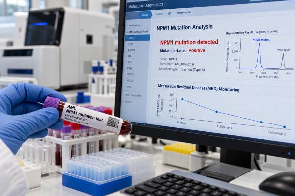
An NPM1 mutation test looks for acquired changes in the NPM1 gene that are especially important in acute myeloid leukemia (AML). NPM1-mutated AML is a genetically defined form of AML, and the result can influence diagnosis, risk assessment, treatment planning, and measurable residual disease (MRD) monitoring. A positive result does not have one fixed meaning: prognosis depends on the full leukemia profile, including cytogenetics, other gene mutations, response to treatment, age, health, and MRD findings over time. NPM1 is also unusually useful after treatment because the leukemia-specific mutation can often be tracked at very low levels with sensitive molecular testing. This makes the marker valuable not only at diagnosis but also during remission, when rising or persistent NPM1 may signal residual leukemia before blood counts or symptoms change. The most useful interpretation therefore combines the initial mutation result with co-mutations and serial MRD measurements rather than reading a single number in isolation.
- A positive NPM1 mutation usually supports a diagnosis of NPM1-mutated AML when interpreted with the blood, bone marrow, morphology, and other molecular findings.
- NPM1-mutated AML is often favorable-risk in modern AML systems, but not automatically: adverse cytogenetics, certain co-mutations, and persistent MRD can change the risk picture.
- NPM1 MRD is commonly measured by sensitive molecular methods after treatment because falling, clearing, persistent, or rising mutation levels can predict relapse risk.
- A negative NPM1 result does not rule out AML. It means this particular molecular marker was not detected and other genetic and cytogenetic tests remain important.
- The most meaningful result is the trend. A baseline NPM1 level at diagnosis allows later tests to show how deeply treatment has reduced the leukemia clone.
Table of Contents
- What the NPM1 Test Detects
- When and How NPM1 Testing Is Done
- How to Interpret NPM1 Results
- NPM1 and AML Prognosis
- NPM1 MRD Monitoring
- How NPM1 Results Affect Treatment Decisions
- Important Limitations and Next Steps
What the NPM1 Test Detects
The NPM1 test detects mutations in nucleophosmin 1, a gene on chromosome 5 that helps regulate several cell functions, including ribosome production, genome stability, and movement of proteins between the nucleus and cytoplasm. In AML, NPM1 mutations typically alter the end of the gene and cause the abnormal NPM1 protein to accumulate in the cytoplasm of leukemia cells.
NPM1 mutations are among the most common acquired genetic abnormalities in adult AML. They occur particularly often in AML with a normal-appearing chromosome pattern. The mutation is usually somatic, meaning it developed in the leukemia cells and was not inherited from a parent. Rare hereditary NPM1 disorders are a different issue and are not what routine AML NPM1 testing is designed to detect.
Modern disease classifications recognize AML with mutated NPM1 as a distinct molecular entity. That matters because the molecular finding can help define the leukemia even when the percentage of blasts in the bone marrow is lower than the classic 20% threshold used for some other AML diagnoses. The full diagnostic rules differ somewhat between classification systems, so a hematopathologist interprets the NPM1 result together with morphology, blast percentage, clinical findings, and other genetics.
NPM1 is not the same as an AML screening test. A positive result strongly supports a clonal myeloid malignancy in the correct context, but testing is usually ordered after an abnormal complete blood count, suspicious blood smear, bone marrow findings, or an established concern for AML. A broader hematologic cancer biomarker evaluation may include cytogenetics, fluorescence in situ hybridization, flow cytometry, and a multigene sequencing panel in addition to NPM1.
When and How NPM1 Testing Is Done
NPM1 testing is usually performed at the time AML is first diagnosed. It may also be repeated after treatment to measure MRD, at suspected relapse, or when a previously treated patient develops new blood-count abnormalities.
The sample is often bone marrow aspirate, because marrow generally contains the highest concentration of leukemia cells at diagnosis. Peripheral blood can also be used when enough circulating blasts are present, and blood is often convenient for serial MRD monitoring. The laboratory report should specify the specimen because a result from marrow and a result from blood are not always directly interchangeable.
Several laboratory methods can detect NPM1 mutations:
| Method | Typical role | Main strength | Main limitation |
|---|---|---|---|
| Next-generation sequencing (NGS) | Diagnosis and broad mutation profiling | Finds NPM1 along with many other AML mutations | Routine panels may not be sensitive enough for very low MRD |
| RT-qPCR | MRD monitoring for common NPM1 mutation types | Very sensitive and quantitative | Requires a validated target matching the patient’s mutation |
| Digital PCR | Highly sensitive molecular quantification | Precise detection at low levels | Availability and assay design vary by laboratory |
| Targeted high-sensitivity NGS | MRD in specialized laboratories | Can track molecular disease at great depth | Technical error suppression and interpretation are critical |
At diagnosis, NPM1 should not be interpreted alone. Important companion tests include karyotyping, FISH, and mutation analysis for genes that influence AML classification and risk. One common example is the FLT3 mutation test. Depending on the panel and clinical setting, testing may also include IDH1, IDH2, CEBPA, TP53, and myelodysplasia-related genes.
No special fasting is usually needed for molecular NPM1 testing. Preparation is instead determined by how the specimen is collected. A blood draw needs little preparation, while a bone marrow aspiration may involve local anesthesia, sedation in some settings, and instructions about medications or eating if sedation is planned.
How to Interpret NPM1 Results
A report may say NPM1 mutation detected, positive, pathogenic variant identified, or not detected. Some reports also give a variant allele frequency (VAF), transcript level, normalized copy number, or log reduction from baseline.
Positive at diagnosis
A positive result means the laboratory found an NPM1 mutation in the tested sample. In a person with compatible marrow findings, this can establish a genetically defined AML subtype. The report may describe the exact DNA change and protein effect or identify a mutation type, such as the common NPM1 type A variant.
The VAF is the fraction of sequencing reads carrying the mutation. For example, a VAF around 40% does not mean that exactly 40% of marrow cells are leukemia cells. VAF is affected by tumor purity, copy number, normal cells mixed into the sample, assay performance, and the genetics of the clone. It is useful molecular information, but it is not a direct substitute for blast percentage.
Negative at diagnosis
A negative test means the assay did not detect an NPM1 mutation above its validated detection limit. It does not exclude AML. Many AML cases are NPM1-wild type and are defined or stratified by other abnormalities. A negative result should therefore be read alongside the complete molecular and cytogenetic workup.
Low-level positive after treatment
After treatment, even a very small positive signal may be clinically meaningful if it is confirmed and represents the same NPM1 mutation that was present at diagnosis. The exact importance depends on the assay, time point, tissue source, degree of reduction from baseline, and whether the signal is stable, falling, or rising.
A single low result near the laboratory’s detection limit may need confirmation. Sample quality, low RNA quantity, and technical variability can affect molecular measurements. A hematology team will often repeat the test rather than make a major treatment decision from one borderline value.
NPM1 and AML Prognosis
NPM1 mutation status is an important part of AML risk classification, but the simple rule “NPM1 positive equals good prognosis” is incomplete.
Under the 2022 European LeukemiaNet framework, NPM1-mutated AML without adverse-risk genetic lesions is generally placed in the favorable-risk group. However, the overall genetic context matters. Adverse cytogenetic abnormalities can outweigh the usual favorable association of NPM1. Co-mutations can also influence biology, treatment sensitivity, and relapse risk.
FLT3 is a particularly important partner test. Earlier risk systems subdivided NPM1-mutated AML according to the FLT3-ITD allelic ratio. The 2022 ELN system no longer uses the FLT3-ITD allelic ratio in the same way, and FLT3-ITD is classified as intermediate risk unless another feature changes the category. This is one reason older explanations of NPM1 prognosis may not match a current report.
Other mutations can add nuance. IDH1 and IDH2 mutations frequently co-occur with NPM1, and targeted drugs may be relevant when those variants are present. The IDH1 mutation result and IDH2 mutation result therefore have both biologic and treatment implications in selected patients.
Most importantly, response to treatment can refine the initial risk estimate. A patient who starts in a favorable genetic group but remains NPM1-MRD positive after therapy may face a higher relapse risk than the diagnostic category alone suggests. Conversely, deep molecular clearance is reassuring, although it never guarantees that relapse cannot occur.
Age, fitness, treatment regimen, transplant eligibility, prior blood disorders, therapy-related disease, and the presence of other high-risk abnormalities also shape prognosis. NPM1 is one powerful variable in a larger clinical model.
NPM1 MRD Monitoring
NPM1 is one of the best-established molecular MRD markers in AML because the mutation is usually stable enough to track and can be measured with assays far more sensitive than routine microscopy.
MRD stands for measurable residual disease: leukemia that remains below the level that can be reliably recognized by standard morphology. A marrow may look like a complete remission under the microscope while molecular testing still detects NPM1-mutated cells. That is why an NPM1 MRD result can add information beyond blast count and blood-cell recovery.
The general pattern is straightforward:
- Large fall from baseline: suggests a strong molecular response.
- Undetectable result: indicates disease is below the assay’s detection limit and is usually associated with a lower relapse risk than persistent positivity.
- Persistent positivity: may indicate residual leukemia and a higher risk of relapse, particularly when levels do not continue to decline.
- Molecular conversion from negative to positive: raises concern for molecular recurrence.
- Rising serial levels: are more concerning than a single isolated low positive result and may precede overt hematologic relapse.
Guidelines recommend molecular MRD assessment at defined treatment milestones in NPM1-mutated AML. Exact schedules differ with treatment strategy and local practice. Testing commonly occurs after induction or early consolidation, after completion of therapy, before or after allogeneic transplant in selected patients, and during follow-up when the result will affect management.
The specimen matters. Bone marrow can be more sensitive at some time points, while peripheral blood is easier to obtain frequently and can provide useful serial information. A laboratory may report MRD as a ratio of NPM1-mutant transcripts to a control gene, a percentage, copies per standardized amount of RNA, or a log change from diagnosis. Because reporting systems vary, it is safer to compare serial values from the same laboratory and assay whenever possible.
Molecular MRD complements, rather than automatically replaces, a broader AML MRD assessment using flow cytometry, PCR, or NGS. Discordant results can occur, and the clinical team may integrate more than one method.
How NPM1 Results Affect Treatment Decisions
At diagnosis, the NPM1 result contributes to the decision framework rather than dictating one treatment by itself. Clinicians combine the molecular profile with age, organ function, performance status, AML subtype, treatment goals, and eligibility for intensive therapy or transplant.
For a fit patient with newly diagnosed NPM1-mutated AML, treatment may include intensive induction therapy followed by consolidation. For older or less fit patients, lower-intensity regimens such as a hypomethylating agent plus venetoclax may be used. The presence of an actionable co-mutation can create additional options. Treatment recommendations evolve quickly, so a mutation report should be linked to the current clinical plan rather than an older risk label.
NPM1 MRD can be particularly important when deciding how much post-remission therapy is needed. In some patients, persistent molecular disease strengthens the case for additional therapy or allogeneic hematopoietic cell transplantation. In others, deep MRD clearance may support avoiding transplant in first remission when the broader risk profile is favorable. The decision is individualized because transplant can reduce relapse risk but introduces major short- and long-term risks of its own.
The marker is also becoming increasingly relevant to targeted therapy. NPM1-mutated AML depends on HOX/MEIS-related transcriptional programs, and menin inhibitors target an interaction that helps sustain this leukemia biology. As new drugs and combinations enter practice, the exact mutation profile can influence trial eligibility and treatment selection.
If relapse is suspected, repeating the molecular panel is useful because AML can evolve. The original NPM1 mutation is often still present, but additional mutations can emerge or change in proportion. Treatment planning at relapse should therefore use current disease biology rather than relying only on the diagnostic specimen.
Important Limitations and Next Steps
NPM1 testing is highly informative, but several common interpretation mistakes can lead to confusion.
First, do not compare raw numbers from different assays as though they were the same scale. A VAF from diagnostic DNA sequencing is not equivalent to an RNA transcript ratio from RT-qPCR. Likewise, MRD percentages from two laboratories may use different controls, calibration systems, and sensitivities.
Second, “not detected” does not mean zero leukemia cells exist. Every assay has a limit of detection. An undetectable result means the target was below that limit in that particular specimen.
Third, NPM1 should not be used as a stand-alone inherited cancer test. A typical AML-associated NPM1 mutation is acquired in the leukemia clone. If the history suggests an inherited myeloid malignancy syndrome, germline testing uses a different strategy and often a non-blood tissue source.
Fourth, the NPM1 result must be distinguished from other mutations that can persist in remission as age-related clonal hematopoiesis. Mutations in genes such as DNMT3A, TET2, and ASXL1 may remain detectable after successful AML treatment and do not always represent the same relapse risk as persistent NPM1. A DNMT3A mutation, for example, may need a different MRD interpretation from NPM1.
Useful questions to ask about an NPM1 report include:
- Was NPM1 detected at diagnosis, and what exact variant was found?
- Were FLT3, cytogenetics, and myelodysplasia-related genes tested at the same time?
- What is the laboratory’s detection limit for the MRD assay?
- Is the current sample blood or bone marrow?
- How does the current level compare with the diagnostic baseline and the previous measurement?
- Does this MRD result change the recommended consolidation, transplant plan, or monitoring schedule?
A new fever, bleeding, severe weakness, shortness of breath, rapidly worsening bruising, or other acute symptoms in a person with AML should be assessed promptly rather than waiting for the next molecular test. NPM1 is a powerful marker of leukemia biology, but clinical status and blood counts remain essential parts of follow-up.
A useful way to review serial NPM1 results is to keep the specimen type and reporting scale consistent. Blood and bone marrow can show different transcript levels, and laboratories may report NPM1 relative to a control gene or with their own validated log-reduction approach. A result should therefore be compared with prior results from the same laboratory whenever possible rather than with an unrelated numerical cutoff from another center.
Timing also changes meaning. Detectable NPM1 immediately after an early treatment cycle is not interpreted the same way as molecular persistence after consolidation or molecular reappearance during remission. Clinicians look at the depth and direction of change, treatment phase, blood-count recovery, and whether the result is confirmed in a repeat sample. A small isolated fluctuation near the assay limit may warrant confirmation, whereas a reproducible rising molecular signal can trigger closer evaluation for impending hematologic relapse.
References
- Diagnosis and management of AML in adults: 2022 recommendations from an international expert panel on behalf of the ELN 2022 (Guideline)
- 2021 Update on MRD in acute myeloid leukemia: a consensus document from the European LeukemiaNet MRD Working Party 2021 (Position Statement)
- Criteria for Diagnosis and Molecular Monitoring of NPM1-Mutated AML 2023 (Review)
- NPM1-Mutated Acute Myeloid Leukemia: Recent Developments and Open Questions 2023 (Review)
- Recent advances in AML with mutated NPM1 2024 (Review)
- NPM1 Measurable Residual Disease: A Narrative Review 2025 (Review)
Disclaimer
This article is for general education and does not replace interpretation by a hematologist, oncologist, or molecular pathologist. NPM1 results must be interpreted with the full AML genetic profile, treatment history, specimen type, assay sensitivity, and MRD trend. Do not change treatment or transplant plans based on a mutation result without discussing it with the treating leukemia team.





