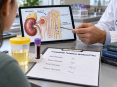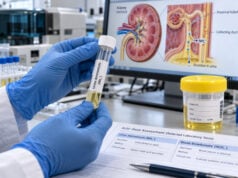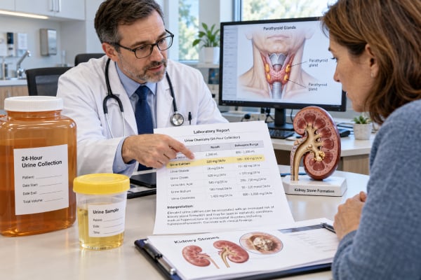
A urine calcium test measures how much calcium is being excreted by the kidneys. It is most often used to evaluate recurrent kidney stones, investigate unusually high or low blood calcium, or help distinguish causes of abnormal parathyroid hormone (PTH) results. Testing may use a spot urine calcium-to-creatinine ratio, but a 24-hour collection is usually more informative when the question is kidney stone risk or total daily calcium loss. A high result is called hypercalciuria and can occur with high sodium intake, primary hyperparathyroidism, idiopathic hypercalciuria, some medicines, or other disorders. A low result may reflect low calcium intake, reduced intestinal absorption, kidney dysfunction, thiazide treatment, or familial hypocalciuric hypercalcemia. The number cannot be interpreted by itself. Collection quality, diet, sodium intake, blood calcium, PTH, kidney function, urine volume, and other stone-risk measurements all affect what the result means.
- A 24-hour urine calcium test is usually preferred for kidney stone evaluation because it measures total calcium excretion over a full day.
- High urine calcium increases calcium-stone risk, but it does not identify the cause by itself. Sodium intake, PTH status, medicines, and genetics all matter.
- Low urine calcium is not always reassuring. It can occur with low intake, malabsorption, chronic kidney disease, thiazide use, or familial hypocalciuric hypercalcemia.
- Primary hyperparathyroidism is diagnosed mainly from blood calcium and PTH, not urine calcium alone. Urine testing helps assess renal involvement and distinguish mimics such as familial hypocalciuric hypercalcemia.
- Collection quality matters. A missed void, unusually high or low diet intake, or an incomplete 24-hour collection can make the result misleading.
Table of Contents
- What a Urine Calcium Test Measures
- Normal, High, and Low Urine Calcium Results
- Causes of High Urine Calcium
- Causes of Low Urine Calcium
- Kidney Stones, PTH, and Hyperparathyroidism
- Collection and Factors That Change Results
- Follow-Up, Treatment, and When to Seek Care
What a Urine Calcium Test Measures
Calcium filtered by the kidneys is mostly reabsorbed back into the blood. Only a small fraction is normally lost in urine. Urine calcium testing measures that excreted fraction and can reveal whether calcium handling is contributing to kidney stones or whether the kidney response fits a blood-calcium disorder.
There are two common approaches.
24-hour urine calcium
A 24-hour collection measures the total mass of calcium excreted during a full day, usually in mg/day or mmol/day. This is the standard approach for metabolic evaluation of many people with recurrent calcium stones. It is often part of a broader 24-hour urine kidney stone risk test that also measures urine volume, sodium, oxalate, citrate, uric acid, pH, and creatinine.
The result answers a practical question: how much calcium reached the urine over the day under the person’s usual conditions? Because stone formation depends on both solute load and urine concentration, clinicians interpret calcium together with urine volume and other constituents rather than as an isolated value.
Spot urine calcium-to-creatinine ratio
A spot sample can report urine calcium concentration alone, but concentration changes markedly with hydration. Dividing urine calcium by urine creatinine partly corrects for dilution. The result may be reported as mg/mg, mmol/mmol, or another laboratory-specific unit.
Spot ratios are useful in selected situations, especially screening or pediatric evaluation, but they do not perfectly substitute for a full-day collection in adults being evaluated for recurrent stones. Time of day, recent meals, sodium intake, and hydration can change a spot result.
Calcium clearance ratios
When blood calcium is high and PTH is not appropriately suppressed, clinicians may calculate a calcium-to-creatinine clearance ratio (CCCR) using paired blood and urine calcium and creatinine. This is different from a simple urine calcium-to-creatinine ratio. CCCR can help distinguish primary hyperparathyroidism from familial hypocalciuric hypercalcemia, although overlap occurs and genetic or family information may still be needed.
A urine calcium result therefore serves several different purposes. The correct interpretation depends on whether the clinical question is stone risk, hypercalcemia, parathyroid disease, bone/mineral balance, or another kidney problem.
Normal, High, and Low Urine Calcium Results
There is no single universal cutoff that applies to every laboratory and every patient. Reference intervals vary with sex, body size, diet, collection method, and the clinical reason for testing.
For 24-hour urine, older clinical conventions have often described hypercalciuria as more than about 250 mg/day in women or 300 mg/day in men, or more than roughly 4 mg/kg/day. Current primary hyperparathyroidism guidance also uses sex-specific urinary calcium thresholds when considering renal involvement. However, kidney stone risk rises continuously with urine calcium rather than suddenly beginning at one cutoff. A value below a traditional threshold can still matter when urine volume is low or other risk factors are present.
To convert calcium units, 1 mmol of calcium is about 40 mg. Thus, 250 mg/day is approximately 6.25 mmol/day and 300 mg/day is about 7.5 mmol/day.
| Pattern | Possible meaning | What usually needs to be checked |
|---|---|---|
| High | Increased filtered calcium, reduced renal reabsorption, high sodium-associated calcium loss, or idiopathic hypercalciuria | Blood calcium, PTH, sodium intake, medicines, kidney stone profile |
| Within laboratory range | May be appropriate, but stone risk still depends on urine volume and other solutes | Oxalate, citrate, uric acid, pH, sodium, stone composition |
| Low | Low intake or absorption, reduced filtration, thiazide effect, or inherited hypocalciuric state | Blood calcium, PTH, vitamin D, kidney function, diet, medicines |
The laboratory’s own reference interval should be used for the final interpretation. A urine calcium result for kidney stone risk is especially useful when compared with prior collections performed under similar diet and medication conditions.
Why concentration alone can be misleading
A urine calcium concentration in mg/dL or mmol/L depends strongly on how much water is in the sample. Someone who produces 3 liters of urine per day can have the same total calcium excretion as someone who produces 1 liter, yet the concentration will be very different. That is why a timed 24-hour total or a calcium-to-creatinine ratio is usually more interpretable than concentration alone.
Creatinine helps assess the collection
Most 24-hour stone profiles also report urine creatinine. Creatinine excretion is not perfectly fixed, but an unexpectedly low amount can suggest a missed collection, while an unexpectedly high amount can suggest overcollection or unusual muscle mass. Clinicians use it as one clue to collection adequacy rather than as an absolute pass/fail test.
Causes of High Urine Calcium
High urinary calcium, or hypercalciuria, is common among people who form calcium stones. It can occur even when blood calcium is normal.
Idiopathic hypercalciuria
In many stone formers, no single secondary disease explains the calcium loss. This is often called idiopathic hypercalciuria. Genetics, intestinal calcium absorption, kidney tubular handling, bone turnover, and hormonal regulation can all contribute. It is associated with calcium stones and, in some people, lower bone mineral density.
Idiopathic hypercalciuria is a diagnosis made after important secondary causes are considered. It does not mean the result is harmless or that treatment is unnecessary.
High sodium intake
Sodium and calcium handling are linked in the kidney. Higher sodium intake generally increases urinary calcium excretion. A high urine sodium result on the same collection can therefore explain part of an elevated calcium value and is often a practical treatment target. The urine sodium test for kidney stone risk can help estimate whether salt intake is contributing.
Reducing excessive sodium may lower urine calcium and also support blood pressure control. This is different from severely restricting dietary calcium, which can sometimes increase intestinal oxalate absorption and worsen calcium oxalate stone risk.
Primary hyperparathyroidism
PTH raises blood calcium by affecting bone, kidney calcium handling, and vitamin D activation. In primary hyperparathyroidism, blood calcium is usually high and PTH is elevated or inappropriately normal rather than suppressed.
The higher filtered calcium load can increase urine calcium, although not every patient with primary hyperparathyroidism has marked hypercalciuria. This is why urine calcium cannot diagnose or exclude the disorder by itself.
Other causes
Other possible contributors include:
- excessive vitamin D or increased vitamin D activation in some granulomatous diseases;
- high calcium supplement exposure in susceptible people;
- prolonged immobilization or high bone resorption;
- hyperthyroidism;
- renal phosphate-wasting disorders;
- loop diuretics, which increase calcium excretion;
- some inherited tubular disorders;
- very high animal-protein and sodium intake patterns.
When urine calcium is high, the important distinction is whether the cause is a modifiable intake pattern, a medication effect, an endocrine disorder, a renal tubular problem, or idiopathic hypercalciuria. Blood tests and the rest of the urine profile narrow that differential.
Causes of Low Urine Calcium
Low urinary calcium is called hypocalciuria. It can be appropriate or abnormal depending on the blood calcium and clinical setting.
Low calcium intake or reduced absorption
Low dietary calcium, vitamin D deficiency, celiac disease, inflammatory intestinal disease, bariatric surgery, and other malabsorption states can reduce the amount of calcium absorbed from the gut and therefore reduce urine calcium.
A low urine value should not automatically prompt calcium supplementation without understanding the reason. The blood calcium, PTH, vitamin D status, diet, and stone type all matter.
Chronic kidney disease
As kidney function falls, filtered calcium load and mineral metabolism change. People with chronic kidney disease may have low urine calcium even when PTH is elevated. Interpretation should therefore include serum creatinine and estimated glomerular filtration rate (eGFR).
Thiazide diuretics
Thiazide and thiazide-like diuretics generally lower urinary calcium by increasing calcium reabsorption in the distal nephron and through related sodium-volume effects. If a test is being used to evaluate baseline calcium excretion, current thiazide use can substantially change the result.
Thiazides have long been used in recurrent calcium stone prevention, but recent trials and reviews have made the benefit-risk picture more nuanced. A fall in urine calcium can show a physiologic drug effect, but the decision to use or increase a thiazide also depends on stone recurrence, blood pressure, potassium, glucose, uric acid, and adverse effects.
Familial hypocalciuric hypercalcemia
Familial hypocalciuric hypercalcemia (FHH) is an inherited disorder, usually involving altered calcium-sensing receptor signaling. It typically causes lifelong mild hypercalcemia with relatively low urinary calcium. PTH may be normal or mildly elevated.
FHH matters because it can resemble primary hyperparathyroidism but usually does not improve with parathyroid surgery. A low CCCR can support FHH, but overlap occurs, especially with vitamin D deficiency, low calcium intake, kidney disease, or thiazide use. Family history, prior calcium results, and sometimes genetic testing are needed.
Kidney Stones, PTH, and Hyperparathyroidism
Most calcium-containing stones form because urine becomes supersaturated with calcium oxalate, calcium phosphate, or both. Urinary calcium is one important part of that chemistry, but it is not the whole story.
Stone risk depends on the complete urine environment
A person with 300 mg/day of urine calcium and 3 liters of urine may have a different supersaturation profile from someone with the same calcium excretion and only 1 liter of urine. Citrate binds calcium and can reduce crystal formation, while oxalate strongly influences calcium oxalate supersaturation. Urine pH changes the balance between calcium oxalate and calcium phosphate risk.
For recurrent stones, clinicians therefore often review:
- total urine volume;
- calcium;
- sodium;
- oxalate;
- citrate;
- uric acid;
- pH;
- creatinine and collection adequacy;
- calculated supersaturation indices.
A urine oxalate test and urine citrate test can be as important as calcium in explaining why stones recur.
How PTH testing fits
If blood calcium is repeatedly elevated, PTH should be measured. In non-parathyroid causes of hypercalcemia, PTH is usually suppressed. In primary hyperparathyroidism, PTH is elevated or inappropriately normal for the high blood calcium.
The parathyroid hormone (PTH) test is therefore central to the diagnosis. Urine calcium has supporting roles: assessing stone-related renal involvement, identifying substantial calcium loss, and helping separate primary hyperparathyroidism from FHH.
Current international primary hyperparathyroidism guidance considers renal findings, including nephrolithiasis, nephrocalcinosis, reduced kidney function, and hypercalciuria, when evaluating patients. Imaging and the full biochemical picture remain important because urine calcium alone does not show whether stones are already present.
A practical pattern comparison
High blood calcium + non-suppressed PTH + normal/high urine calcium: primary hyperparathyroidism becomes more likely, although the diagnosis uses the full clinical picture.
High blood calcium + non-suppressed PTH + very low urine calcium/low CCCR: FHH becomes an important possibility, but confounders must be excluded.
Normal blood calcium + high urine calcium + recurrent calcium stones: idiopathic hypercalciuria, high sodium intake, or another secondary cause may be considered.
Low or normal urine calcium + recurrent stones: do not stop the evaluation. Low urine volume, high oxalate, low citrate, abnormal pH, uric acid, infection, or anatomic factors may still drive stone formation.
Collection and Factors That Change Results
A 24-hour urine calcium result is only as reliable as the collection.
How a 24-hour collection is usually done
On the starting morning, urinate into the toilet and record the time. After that, collect every urine sample for the next 24 hours, including the final sample at the same time the following morning. Follow the laboratory’s instructions for the collection container, preservative, and refrigeration.
If a substantial sample is missed, the collection may need to be repeated. Do not add extra hours to compensate. A 24-hour urine calcium test should represent a defined period.
For stone prevention, many clinicians want the patient eating and drinking in a reasonably typical way so the result reflects usual exposure. If the purpose is to test a specific treatment, the collection may instead be performed after a planned change.
Diet can change calcium excretion quickly
Urinary calcium is influenced by:
- sodium intake;
- calcium intake;
- animal-protein intake;
- fluid intake and urine volume;
- vitamin D and calcium supplements;
- fasting or unusual dietary patterns.
Do not intentionally restrict calcium before a stone evaluation unless directed. Normal dietary calcium with meals can bind oxalate in the gut. Very low calcium intake can lower urine calcium while increasing oxalate absorption, which may produce a misleadingly “better” calcium number without lowering stone risk.
Medicines and illness matter
Thiazides lower urine calcium, while loop diuretics can raise it. Corticosteroids, vitamin D preparations, calcium supplements, lithium, and several other medicines can alter calcium or PTH physiology. Never stop a prescribed medicine solely to change the test unless the ordering clinician instructs you.
Dehydration, vomiting, diarrhea, IV fluids, acute kidney injury, and major changes in activity can also make a collection unrepresentative. If the result does not fit the clinical picture, repeating the test under more stable conditions is often more useful than overinterpreting a single number.
Check the units
A common source of confusion is comparing a spot concentration, a spot calcium-to-creatinine ratio, and a 24-hour total as if they were the same test. They are not. Always confirm whether the result is mg/dL, mmol/L, mg/g creatinine, mg/mg creatinine, mmol/mmol, mg/day, or mmol/day before applying a cutoff.
Follow-Up, Treatment, and When to Seek Care
Follow-up depends on why the test was ordered and what else is abnormal.
For recurrent calcium stones, clinicians often compare urine calcium with stone composition and the full 24-hour urine profile. Common first measures include increasing fluid intake enough to produce a higher urine volume and reducing excessive sodium. Dietary calcium is usually maintained at an appropriate level rather than severely restricted. Treatment can also target low citrate, high oxalate, high uric acid, or abnormal urine pH.
If hypercalciuria persists, clinicians look for secondary causes. Blood testing may include calcium, albumin, phosphorus, creatinine, bicarbonate, PTH, and 25-hydroxyvitamin D. The pattern determines whether endocrinology, nephrology, or urology evaluation is most useful.
Drug treatment is individualized. Thiazide or thiazide-like diuretics can lower urine calcium, but contemporary evidence shows that lower urine calcium does not automatically guarantee fewer symptomatic stones for every patient. Blood pressure, potassium, sodium intake, glucose, uric acid, kidney function, and adverse effects should be monitored. Treatment decisions are based on recurrence risk and the whole metabolic profile rather than a urine calcium target alone.
For primary hyperparathyroidism, treatment decisions use blood calcium, age, bone density, kidney function, stones or nephrocalcinosis, urine calcium, symptoms, and patient preferences. Surgery is definitive treatment when indicated. Urine calcium is part of this assessment but does not determine the diagnosis or operation on its own.
For low urine calcium with hypercalcemia, clinicians should review calcium intake, vitamin D, kidney function, thiazide use, and the CCCR before concluding that FHH is present. If the biochemical pattern and family history remain suggestive, genetic testing may be considered.
Seek urgent medical care for severe flank pain with fever or chills, inability to pass urine, persistent vomiting with dehydration, confusion, severe weakness, or symptoms associated with markedly abnormal blood calcium. Kidney stones plus fever can indicate an obstructed infected urinary system, which requires prompt evaluation.
For nonurgent abnormalities, the most useful next step is often a planned repeat or complementary test rather than reacting to one isolated value. A well-collected urine result interpreted alongside blood calcium, PTH, kidney function, diet, medication exposure, and stone history gives a much clearer answer.
References
- Evaluation and Management of Primary Hyperparathyroidism: Summary Statement and Guidelines from the Fifth International Workshop 2022 (Guideline)
- Idiopathic Hypercalciuria: A Comprehensive Review of Clinical Insights and Management Strategies 2025 (Review)
- Thiazides for kidney stone recurrence prevention 2024 (Review)
- Thiazide Dose, Urine Calcium, and Symptomatic Kidney Stone Events 2024 (Cohort Study)
- Baseline Urinary Calcium and the Efficacy of Thiazide Diuretics for Kidney Stone Prevention. 2026 (Cohort Study)
- Contrasting Response of Urine Stone Risk to Medical Treatment in Calcium Oxalate versus Calcium Phosphate Stone Formers 2024 (Cohort Study)
Disclaimer
Urine calcium results must be interpreted with blood calcium, PTH, kidney function, diet, medication use, and the reason for testing. A single high or low value does not diagnose a parathyroid disorder or determine kidney stone treatment. Seek medical care promptly for severe stone symptoms, fever with urinary obstruction symptoms, or symptoms of severe calcium imbalance.